Recent Roadwork at the Intersection of Science and Art

It seems it’s been eons since my last post to Evolutionary Routes; the blog was well on its way to becoming a great case study in fossilization. In light of my insomnolent work schedule these days (on which I blame my tardy posting habits, and I’m sticking to my story), I have decided to try a new strategy to increase the frequency of posts: shorter, bite-sized blog posts. Hopefully these will also be less arduous to read, so there’s something in it for you too as the reader.

Let me kick off with a shameless self-plug that nonetheless explains why posts here have been so scarce lately. As much as I love it nearly to death, I’ve been up to my eyeballs in meeting deadlines for my work. This work consists of natural history art projects involving museum exhibits, books, coin designs and scientific research press release images featuring dinosaurs, all things prehistoric and some extant natural history thrown in for good measure.

A couple of news items from my work, relating evolutionary biology to art stand out in particular.

Paleoart Book

1554470_10151863089800583_1082699473_n

In May, a book exclusively featuring my paleoart was released by Titan Books. Entitled “The Paleoart of Julius Csotonyi: Dinosaurs, Sabre-tooths and Beyond” by Julius Csotonyi and Steve White, this 156-page coffee table book provides a pretty comprehensive exhibit of my artwork that focuses on the use of paleontological science to restore the life appearances of not only dinosaurs, but also entire prehistoric ecosystems. Having been more than a year in the making, its content is mostly artwork, but also contains written contributions from about 20 paleontologists with whom I’ve collaborated to restore prehistoric life. Should you be interested in ordering it, the book is available through most major book sellers online; just search for “The Paleoart of Julius Csotonyi”.

Paleoart Website

Csotonyi-dot-com_01_Acheroraptor

More recently, my natural history art website, csotonyi.com, which has been in developmental limbo for longer than Evolutionary Routes, has also received a much needed revamping. My wife, Alexandra Lefort, has applied her web design wizardy to it to come up with a sleek new look, and we’ve added to it a lot of new pieces of my artwork resulting from close collaboration with paleontologists and museum curators. There’s even a new art blog (because I don’t have enough writing to do on a science blog that I maintain proficiently). My intent is to share mainly art and illustration-themed thoughts there, focusing on how I complete restorations, instead of the more purely science-related topics here.

Both resources focus very heavily on visual restoration of evolutionary biology, and whereas many of the pieces on the website also appear in the book, the book additionally contains reproductions of museum murals that appear only within its pages or on the walls of the host museums. The website will be updated each time I publish a new piece of artwork. Enjoy!

Posted in Uncategorized | 5 Comments

Why do two dragons sleep soundly?

The second specimen of the small Chinese Cretaceous troodontid dinosaur, Mei long, curled up under a volcanic ash fall, asphyxiated either in its sleep or in a defensive posture. Image source: Julius T. Csotonyi.

The little crow-sized troodontid dinosaur, Mei long (meaning “soundly sleeping dragon”) was more aptly named than Xu and Norell (2004) originally realized. Now, Chunling Gao and colleages (2012) have reported their discovery of a second fully articulated specimen preserved in the posture characteristically adopted by many modern birds while they sleep: neck curved around sideways, head tucked in behind a wing, legs folded under body and (something of which modern birds are incapable, with their truncated pygostyle) long tail wrapped around the body. The only significant difference in posture between the two animals is that the new one’s (DNHM D2154) neck is bent in the opposite direction to that in the first specimen (IVPP V12733). Xu and Norell (2004) had suggested that as in modern birds, this compact body configuration minimizes the ratio of surface area to volume, thereby enhancing body heat retention, which is a significant challenge to such small homeothermic (warm blooded) animals.

Despite the fact that these fossils were found in the volcaniclastic (volcanic ash deposit) sediments of the Yixian formation, which facilitate extremely fine preservation, fossilization is a very spotty process (only two such beautiful Mei specimens are known so far). So it seems statistically unlikely that both specimens (and a partial Sinornithoides) would be found in this sleeping posture unless death and the posture were somehow related. But how?

Gao et al. (2012) point out that not all the victims of Pompei (who were similarly covered by ash as Mei) display the hyperflexion (clenching) of muscles that results from death by high temperature ash falls and fire. Therefore, (assuming that theropods such as Mei display similar neuromuscular responses to heat as do mammals) it isn’t unreasonable to surmise that the specimens of Mei died by asphyxiation from toxic volcanic gases while sleeping instead of being roasted. In this scenario, the volcanic eruptions that killed and buried the animals may have occurred during the sleeping period, and therefore represents a kind of snapshot of ‘normal’ life in this prehistoric ecosystem.

Alternatively, Gao et al. (2012) have suggested that perhaps the preserved brooding posture of Mei represents either (1) sheltering behaviour within a burrow (which may have constrained the position of the body and prevented the kind of opisthotonic neck-twisted-up-over-back posture that characterizes many dinosaur skeletons and which has been proposed to results from either a neurolomuscular response to toxins or agony, or postmortem contraction of neck ligaments), or (2) a defensive postural response to volcanic events such as ash fall. In this last case, the volcanic event may have directly influenced the preserved postures of some of the fossils that are found in its resulting sediments. In the sketch above, I have chosen to illustrate Mei curled up under a relatively cooler ash fall, and the hapless creature has been asphyxiated, either in its sleep or in a defensive posture.

The multiplicity of possible explanations for the brooding posture of Mei and the likelihood of the fossilization process to influence the behaviour of the animals that it preserves underscores the daunting and complex task facing paleontologists who strive to piece together the puzzle of prehistoric ecosystems from relatively few pieces left to them sometimes by catastrophic events.

References

Gao C., Morschhauser E.M., Varricchio D.J., Liu J., Zhao B. (2012). A Second Soundly Sleeping Dragon: New Anatomical Details of the Chinese Troodontid Mei long with Implications for Phylogeny and Taphonomy. PLoS One. 7(9): e45203.

Xu X., Norell M., (2004). A new troodontid dinosaur from China with avian-like sleeping posture. Nature. 431(7010): 838-841.

Posted in Uncategorized | 2 Comments

Sinocalliopteryx and its feathered fast food

What’s so exciting about the recent report by Xing et al. (2012) of the feathered meals of a fuzzy compsognathid dinosaur known as Sinocalliopteryx? I mean, we’ve known for years that dinosaurs ate other dinosaurs, right? To me, this is a fine example of how the investigative power of paleontology is growing as its knowledge base increases, and shows how fossil remains have so any more stories to tell than just the appearance of the animal that left them.

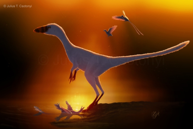
Sinocalliopteryx standing over its Sinornithosaurus prey, as a breeding pair of Confuciusornis take flight behind. Both latter genera have been found as prey of different Sinocalliopteryx individuals. The scene is set on the shores of the lake in which they will ultimately be preserved in ash. Image source: Julius T. Csotonyi

Meet Sinocalliopteryx: Bad news for feathered friends

As you may have read, the news is based on two skeletons of a flightless dinosaur with filamentous plumage, Sinocalliopteryx, from the wonderful Yixian Formation of China, from volcaniclastic lacustrine deposits (volcanic ash transported by wind or water and deposited in a lake — great for preserving fine anatomical details). The first skeleton, designated JMP-V-05-8-01, was the original specimen from which the species was described, and was noted to contain a limb of another theropod in its abdomen even when it was described (Ji et al., 2007). This limb has since been shown to belong to a feathered dromaeosaurid, Sinornithosaurus (Xing et al., 2012). The JMP-V-05-8-01 individual has the exceptionally complete skeleton of which you frequently see photos for this genus. The second skeleton, designated CAGS-IG-T1, is incomplete but the portions that have been recovered contained gut contents that have been diagnosed as the early bird, Confuciusornis, and also part of an ornithischian dinosaur.

Head of Sinocalliopteryx. Image source: Julius T. Csotonyi

What can Sinocalliopteryx fossils teach us beyond just morphology?

Aside from the exquisitely fine state of preservation that the Sinocalliopteryx fossils exhibit, allowing us to know precisely how their bones fitted together and even the appearance of their long filamentous feather-like integument, this kind of composite fossil evidence also reveals how prehistoric ecosystems functioned, and how their component species interacted with each other. This interaction of prehistoric organisms with each other and with their environment, encompassing a field of study known as paleoecology, provides some very important pieces of the puzzle of knowldge.

The way that an animal’s remains are associated with trace fossils (footprints, skin impressins, coprolites — fossilized poop, etc.) and with the fossilized parts of other animals with which they are preserved can reveal how they behaved or interacted with each other. When we consider the fossils from a particular locality, simply enumerating the species that lived contemporaneously is akin to reading the cast of a movie. Knowing something about how they interacted — their paleoecology — is like piecing together the movie’s plot or script. It gives us a much richer snapshot of what life was like in these extinct biological communities.

A lot more can be gleaned from fossils such as these Sinocalliopteryx than merely that dinosaurs ate other dinosaurs, especially when related observations from paleontology are brought to bear on the study. For example:

(1) Lida Xing and colleagues used the shape of the gastrointestinal tract of another phenomenally preserved small theropod, Scipionyx, to help determine how the digested meal of Sinocalliopteryx progressed through the digestive tract of the latter.

(2) The authors noted that the second specimen of Sinocalliopteryx (CAGS-IG-T1) contained a highly pitted and therefore relatively more digested scapula (shoulder blade) of an ornithischian dinosaur, possibly either Psittacosaurus or Yueosaurus. Poor Psittacosaurus — always getting eaten. First, embarrassingly, by a mammal known as Repenomamus (Hu et al., 2005), and now possibly by Sinocalliopteryx. The advanced state of digestion implied that unlike modern predatory birds, which expell bones by regurgitation after consuming their prey, dinosaurs such as Sinocalliopteryx passed bone material right through their digestive system. This has implications on their stomach acidity. The reason that modern birds regurgitate bones is that their stomach is not acidic enough to sufficiently soften ingested bones, making them a hazard to retain. The authors pont out that modern alligators (a sister archosaur group to dinosaurs), by contrast, can make their stomach significantly more acidic (pH of 1.2) by shunting deoxygenated blood to the stomach, allowing them to deal with large boney meals. These observations suggest that Sinocalliopteryx (and other dinosaurs, based on the boney contents of coprolites) may have possessed a digestive system more similar to that of their crocodilian cousins than the feathery descendents of dinosaurs.

(3) The CAGS-IG-T1 specimen possessed in its abdomen the remains of at least two Confuciusornis. This raised the interesting prospect that Sinocalliopteryx was active and agile enough to hunt flying prey despite being ground-bound itself. Of course, the authors admit that it might have simply scavenged dead birds — which are a whole lot easier to catch — but they also pointed out that the probability of a scavenger finding not one but two downed individuals of the same species in rapid succession (both Confusiusornis specimens were at a similarly low early stage of digestion) seems low (unless, perhaps, they were both the victims of a widespread phenomenon, such as volcanic carbon dioxide degassing, but this, too, seems unparsimonious). Add to this the fact that the JMP-V-05-8-01 specimen had fed on the feathered (and possibly flight- or at least gliding-capable) Sinornithosaurus, and the case for a cat-like, bird-hunting, ambush-predator strengthens (Xing et al., 2012).

Male (with long caudal feathers) and female Confuciusornis. Parts of at least two of these early birds were found in the abdomenal region of the CAGS-IG-T1 specimen of Sinocalliopteryx. Like all earliest birds, Confuciusornis possessed three clawed fingers on each hand. Image source: Julius T. Csotonyi

So the Sinocalliopteryx skeletons have, by way of the high-fidelity preservation of the original positions of their components, gut contents and all, taught us about not only their morphology, but also their digestive processes, dietary preferences and probable level of activity and agility. Not bad for some silent bones.

Paleoecological showstoppers

Some other spectacular fossils (just a very short list of myriad others) that have yielded valuable paleoecological insights include the following.

Psittacosaurus (the ornithischian dinosaur whose putative remains were found in the abdomen of one of the Sinocalliopteryx individuals) deserves special mention, for several specimens have provided surprising paleoecological insight. As described above, the badger-like Cretaceous mammal, Repenomamus, was found with the remains of young Psittacosaurus in its abdomen (Hu et al., 2005), illustrating that mammals occasionally reversed the direction of the arrow of predation on dinosaurs.

The Cretaceous mammal Repenomamus consuming Psittacosaurus. Image source: Julius T. Csotonyi

Furthermore, not only did an exquisite specimen of Psittacosaurus from the fine Yixian sediments demonstrate that some ceratopsians possessed long, bristle-like integumentary structures on their tail (Mayr et al., 2002), but a tight group of 34 juveniles were unearthed with a single adult animal in another find (Meng et al., 2004). The ossification of the bones of the young implied that they were juveniles, not embryos, and provided impressive support for parental care in this genus.

Psittacosaurus adult with 34 young, from Yixian Formation, China. Image source: Harry Nguyen (Wikimedia Commons)

A delicately preserved fossil of a small troodontid, Mei, also from the Yixian formation, was curled in a posture typical of modern sleeping birds when it was buried by volcanic ash (Xu & Norell, 2004), suggesting that some avian behaviours may be very old, perhaps originating in their common ancestor with sister theropod groups. Alternatively, such behavioural modes may have evolved independently more than once.

Mei, reconstructed in the sleeping posture in which its remains were found. Image source: Julius T. Csotonyi

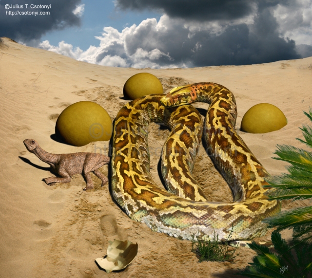
Catching animals in the act of predation is rare, but sometimes death strikes at such moments, and if we are extremely lucky and conditions are just right, they my be fossilized. Examples include a titanosaur nest with eggs, being invaded by the snake Sanajeh, implying that predation by snakes was one of the hazards that faced dinosaur eggs and hatchlings (Wilson et al., 2010).

Reconstruction of the remarkable fossil of the prehistoric snake, Sanajeh, raiding the nest of a titanosaur. Image source: Julius T. Csotonyi

Several fossils of Cipati (a relative of Oviraptor) have been found brooding clutches of eggs (Norell et al., 1995; Clark et al., 1999), which seems to imply that death by sand burial while administering parental care was a disturbingly common fate for this animal.

There’s the beautiful recent find of a pregnant plesiosaur (Polycotylus), which showed us that these marine reptiles shared K-selected viviparous (giving live birth) reproductive life history characteristics with cetaceans (whales) and ichthyosaurs rather than their oviparous (egg laying) reptilian relatives, sea turtles (O’Keefe & Chiappe, 2011). K-selected organisms invest relatively much energy into the production of young, employing a strategy of maximizing the survival of a few young rather than releasing many small young that each have a lower probability of survival. The K-selected strategy is typically used by organisms whose population is relatively stable and often near the carrying capacty (K) of the environment.

Restoration of Polycotylus giving live birth. Image source: Julius T. Csotonyi

Paleoecology and lagerstätten

Inferences about paleoecology do not always demand fully articulated skeletons. For example, bones of hadrosaurs (duckbill), Triceratops and Tyrannosaurus bearing characteristic tooth scrapes can tell us that Tyrannosaurus not only fed on certain groups of herbivores but also exhibited cannibalism (Longrich et al., 2010). However, as the Sinocalliopteryx findings demonstrate, the preservation of some types of behaviour or physiological characteristics do require exceptionally high quality of preservation, because some types of information can only be conveyed by the relative orientation of preserved parts with respect to each other.

Hence, geological formations generated by environments that catalyzed high quality fossilization are crucial to a fuller understanding of how life played out in the distant past. These formations represent the clearest snapshots of the past, as if some giant slapped shut a giant phonebook on nearly an entire ecosystem and pressed it like so many flowers. Such fossil localities that are remarkable for their diversity and/or quality of preservation are known as lagerstätten. Many of the finest examples of how prehistoric organisms interacted with each other come from lagerstätten.

Now, a humbling thought: consider how little of the past we are able to view at this spectacular level of detail. Lagerstätten possessing the quality of preservation of the Yixian lakebeds, which preserve soft tissues, are relatively rare. Some of the best known are: Ediacara Hills (Precambrian, Australia), Chengjiang (Cambrian, China), Burgess Shale (Cambrian, Canada), Wheeler Shale (Cambrian, USA), Much Wenlock Limestone (Silurian, Great Britain), Rhynie Chert (Devonian, Scotland), Cleveland Shale (Devonian, USA), Bear Gulch (Carboniferous, USA), Gres à Voltzia (Triassic, France), Holzmaden (Jurassic, Germany), Solnhofen Limestone (Jurassic, Germany), Jehol Group (Cretaceous, China), Santana Formation (Cretaceous, Brazil), Green River Formation (Eocene, USA) and the Messel Shale (Eocene, Germany).

For a time span of over half a billion years, a list even several times this size would be tear-jerkingly short. Imagine how many fascinating snapshots of time we will never be able to view from the biosphere’s early years because the conditions of fossilization were, as is usually the case, unfavourable for the pristine preservation that is needed to piece together the ecological script. Fossilization is such a finicky process that we should not be surprised to find gaps in the fossil record. It is also why those rare gems of geological formations that we do find are so vitally important to understanding the history of our planet.

References

Clark, J.M., Norell, M.A., & Chiappe, L.M. (1999) An oviraptorid skeleton from the Late Cretaceous of Ukhaa Tolgod, Mongolia, preserved in an avianlike brooding position over an oviraptorid nest. American Museum Novitates, 3265: 36 pp. American Museum of Natural History, New York.

Hu, Y.; Meng, J.; Wang, Y.; Li, C. (2005) Large Mesozoic mammals fed on young dinosaurs. Nature. 433: 149-152.

Ji, S.; Ji, Q.; Lu J.; Yuan, C. (2007) A new giant compsognathid dinosaur with long filamentous integuments from Lower Cretaceous of Northeastern China. Acta Geologica Sinica. 81(1): 8-15.

Longrich, N.R.; Horner, J.R.; Erickson, G.M.; Currie, P.J. (2010) Cannibalism in Tyrannosaurus rex. PLoS ONE 5(10): e13419.

Mayr, Gerald, Peters, D. Stephan, Plodowski, Gerhard & Vogel, Olaf. (2002 Bristle-like integumentary structures at the tail of the horned dinosaur Psittacosaurus. Naturwissenschaften 89: 361–365.

Meng, Q; Liu, J; Varrichio, D.J.; Huang, T.; Gao, C. (2004) Parental care in an ornithischian dinosaur. Nature 431: 145–146.

Norell, M.A., Clark, J.M., Chiappe, L.M., and Dashzeveg, D. (1995) A nesting dinosaur. Nature 378:774-776.

O’Keefe, F.F.; Chiappe, L.M. (2011) Viviparity and K-Selected Life History in a Mesozoic Marine Plesiosaur (Reptilia, Sauropterygia). Science: 333 (6044): 870.

Wilson JA, Mohabey DM, Peters SE, Head JJ. (2010) Predation upon Hatchling Dinosaurs by a New Snake from the Late Cretaceous of India. PLoS Biology. 8(3): e1000322

Xing, L; Bell, P.R.; Persons, W.S. IV; Ji, S.; Miyashita, T.; Burns, M.E.; Ji. Q.; Currie, P.J. (2012) Abdominal contents from two large early Cretaceous compsognathids (Dinosauria: Theropoda) demonstrate feeding on confuciusornithids and dromaeosaurids. PLoS ONE 7(8): e44012.

Xu and Norell, (2004). A new troodontid dinosaur from China with avian-like sleeping posture. Nature, 431(7010): 838-841.

Posted in Uncategorized | 5 Comments

Milestones

On July 1, Evolutionary Routes completed its first tour around the solar system, turning one year old. It is rewarding to watch the readership of the blog increasing, and some thoughtful comments being contributed. A big thank you goes out to all of my readers, and to those who have taken the additional time to leave comments, which are always welcome.

As I commented on a previous post, a large part of my work entails scientific illustration. As a result, some of the first year of blog writing was displaced by the demands of mural illustration projects for museums and publications. I described my production process of the murals for the new Hall of Paleontology at the Houston Museum of Natural Science in a previous post.

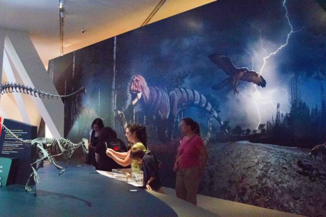
One of the unusual dinosaurs unearthed from Saharan Africa, Ouranosaurus, poses in front of a mural restoration of its Cretaceous environment at the ROM’s Ultimate Dinosaurs: Giants from Gondwana exhibition. Unlike the remaining four murals, this mural is projected onto a screen and makes use of moving imagery, i.e. dynamically changing clouds that I illustrated on a separate layer from the rest of the background. Artwork by Julius Csotonyi; Photograph by Alexandra Lefort.

More recently, another exhibit featuring my paleoart (artwork that endeavors to reconstruct prehistoric subjects) opened at the Royal Ontario Museum (ROM) in Toronto, Ontario, Canada. Ultimate Dinosaurs: Giants from Gondwana, was curated by Dr. David Evans and Dr. Matthew Vavrek. This project focused on the unusual dinosaur fauna of the southern hemisphere, a subject that few northern hemisphere museums have covered, and none to date in Canada to this extent. What’s great about this exhibit from an evolutionary biology standpoint is that it weaves together the fascinating geological and paleontological records that describe how the development and ultimate break-up of the ancient supercontinent of Gondwana affected the evolution of the bizarre assemblage of dinosaurs that are being unearthed in the southern hemisphere. The exhibition winds through a large space, telling the story of prehistoric environments from sites in Argentina, Niger, Madagascar and Patagonia, comparing and contrasting these southern ecosystems to more familiar northern counterparts, such as the Hell Creek formation in North America Most of the dinosaurs featured in the exhibit have only been described within the last ten years. Among the unusual genera (which can be viewed here) displayed in the exhibit are the spined sauropod Amargasaurus, sail-backed Ouranosaurus, crocodile-jawed Suchomimus, enormous sauropod Futalognkosaurus, horned theropods Carnotaurus and Majungasaurus and the enormous predator Giganotosaurus.

Casts of Triassic animals in the ROM’s exhibit stand in front of a mural reconstruction of their Argentinian environment. Animals include Herrerasaurus, Pisanosaurus, Eoraptor and Prestosuchus (the latter not a dinosaur, but a rauisuchian archosaur). Artwork by Julius Csotonyi; Photograph by Alexandra Lefort.

For this project, I created five large murals of dinosaurs and contemporaneous flora and fauna in environmental reconstructions (up to 15 x 150 feet, or 5 x 50 m, in size), as well as seventeen dinosaur vignette illustrations for accompanying information panels. The murals feature mostly full sized restorations of dinosaurs, positioned beside and behind the skeletons, and in the same positions as the skeletal mounts, allowing visitors to compare the skeletal anatomy to the fleshed-out restorations at the same scale.

Visitors examine skeletons from Cretaceous Madagascar. Behind them, a mural features Majungasaurus ambushing a young Rapetosaurus, as a Rahonavis glides off in escape, while a Masiakasaurus toys with a Beelzebufo. Artwork by Julius Csotonyi; Photograph by Alexandra Lefort.

The exhibition also features full sized casts of 17 dinosaurs, produced by Peter May’s renowned team of Research Casting International. Unusual for museums, but becoming more extensively utilized, the exhibit also features augmented reality components, such as rotatable digital tablets that allow users to visualize how the dinosaur would have looked fleshed out and moving, and reactive wall displays that incorporate motion sensors to selectively animate plant and animal components of the murals as visitors approach them. To create these innovative components of the exhibit, layered files of the murals and dinosaur illustrations were used in the generation of animated 3-dimensional models by a team of modellers at Meld Media, and the extraordinary artists Andrey Atuchin and Vlad Konstantinov.

Yours truly playing with one of the swivelling digital tablets installed to allow visitors to see what the mounted skeletons (in this case, Tyrannosaurus) would have looked like in life, fleshed out, with tails whipping and jaws snapping. Artwork by Julius Csotonyi, Andrey Atuchin and Vlad Konstantinov; Photograph by Alexandra Lefort.

Speaking of annual milestones, because July 1 is also Canada’s national birthday, I’ll wish my fellow Canadians a (slightly belated) happy 145th Canada Day. And, since the Fourth of July is just peeking around the corner, I will end this post with a wish for happy national birthday festivities to our neighbors in the U.S.A.

Posted in dinosaurs | Tagged , | 2 Comments

Evolution Powered by Lightning?

In the film derivation of Mary Shelley’s classic, “Frankenstein”, the eccentric doctor floods the body of his chimaeric creation with the energy from a lightning bolt, imparting the monstrous biological assemblage with new life. Pure science fiction?

Well, perhaps not completely.

I’m not referring to the surge of electricity that can sometimes reanimate the heart of a trauma patient. I am also not talking about any discharges of energy that may or may not have catalyzed any of the early chemical reactions on the way to abiogenesis on the primordial earth. I am referring to the potential ability of lightning to accelerate evolution in living soil bacteria, to help living bacteria acquire ‘new life’ in the form of novel physiological abilities.

Horizontal Gene Transfer (HGT)

Recall that in an earlier post (“Evolution’s Usual Suspects: 1. Plagiarizing Wizards“) I described the remarkable ability of many bacteria to integrate into their own chromosome foreign fragments of DNA discarded by other species. This process of horizontal gene transfer (HGT) potentially allows them to acquire physiological traits (e.g. the capacity to break down new compounds for energy) that are encoded on the genes within the recovered fragments. The process could accelerate evolution because once such physiological abilities have evolved once, it provides a shortcut that circumvents the need to repeatedly evolve them in different lineages through lengthy series of mutations.

Graphical representation of horizontal gene transfer. The branched tree-like structure represents the evolutionary lineage (geneological tree) of representatives of earth’s major types of life forms. Sometimes genes can be transferred (horizontal gene transfer) between otherwise distantly related species. This is illustrated by bridges forming between branches of the tree, where genes ‘jump’ from one lineage to another. (Image: Barth F. Smets, Ph.D.)

But how do these bacteria actually take up the DNA fragments from their environment? This is not a trivial problem, and it is at this step that lightning may play a surprising role.

Why is the taking up of DNA a problem? Let’s take a quick refresher of the structure of the bacterial cell envelope, which separates the contents of the bacterium from its environment. If you’re already familiar with the phospholipid bilayer, you can skip to the next section.

The Phospholipid Bilayer — a Refresher

Enter the phospholipid molecule. It looks roughly like a bobby pin, with one head and two parallel tails sticking out of it. The phosphate head is a phosphorous atom with oxygen chemically bonded to it. The lipid tails are mainly long chains of carbon and hydrogen. Together the assembly is known as a phospholipid molecule.

Schematic showing how phospholipid molecules and other components fit into a generalized cell membrane. Image source: Mariana Ruiz (Wikimedia Commons)

Now, recall from chemistry classes on solvents that “like dissolves like”. Water molecules are very polar; i.e. they are relatively more positively charged on one side and relatively more negatively charged on the other. Liquids composed of polar molecules such as water can dissolve other substances that are also relatively polar or charged (such as the aforementioned phosphate head). These water soluble substances are hydrophilic (‘water loving’). But water will not effectively dissolve nonpolar molecules, such as fats, or their subunits, lipids. This includes the lipid tails of the phospholipids.

So, when we try to dissolve phospholipids in water, the hydrophobic (‘water fearing’) lipid tails will clump together, apparently ‘seeking escape’ from the water. This results in a central aggregation of lipid tails all pointing toward each other, with the hydrophilic phosphate heads sticking out all around, arranged as if on the surface of a sphere. These aggregate structures look like little round droplets at extremely high magnification.

What happens if we add lots more phospholipids? Think of what happens when you pull on the ends of a zip-locked bag sealed without any air inside. It spread out flatly, forming a plane composed of two adhering sheets. A sea of phosphate heads form the two outer surfaces of this phospholipid sheet, with the lipid tails hidden between them. A flexible sheet is not a stable structure (think about how your unmade bedsheets’ most stable configuration always appears to be a crumpled mess), so the phospholipid sheet warps and bends until the edges end up meeting. Wherever the edges of this thin phospholipid membrane find each other, they ‘heal’ together, completely hiding the lipids from view of the water, and forming a closed bubble, or vesicle.

We now effectively have a bag composed of a two-layered membrane (a phospholipid bilayer), which is the recipe for holding in the components of a cell — yours, your dog’s your plants’, and yes, those of the bacteria in your gut. There are other rivet-like molecules that cells insert within their phospholipid membranes to strengthen them, and plants, fungi and bacteria perform additional strengthening tricks, such as lining their phospholipid cell membranes with walls composed of cellulose, chitin or peptidoglycan, or adding an extra layer of membrane, but we’ll focus on a simplified cell membrane for now.

“You shall not pass”…Mostly

The phospholipid bilayer is an efficient way of keeping the cell’s components in and much of the environment out. Only certain small molecules may pass through this semi-permeable membrane. In fact, it’s so efficient a barrier that it prevents entry to certain components that the cell needs. So a way must be found to allow entry to desired molecules. Certain charged molecules and large molecules find it especially difficult to pass. DNA is one example.

DNA at Cellular Border Crossings

DNA usually needs not cross the bacterial cell membrane, but in the previous post about HGT, we saw how the integration of foreign DNA fragments could substantially increase the fitness of some bacteria by endowing them with new and useful physiological abilities.

Although many bacteria have evolved special systems of receptors and pores (controlled gateways through the cell membrane) to facilitate uptake of foreign DNA fragments, there are other ways to move DNA across the cell membrane. For example, exposure to certain chemicals, such as calcium chloride, followed by a heat shock, can do this.

Electroporation

In 1982, a team of scientists in Germany [1] found that zapping cells with brief pulses of 80,000 volts of electricity can also elicit DNA uptake. Their objective was to find a way to introduce foreign DNA into cells in order to transform them, meaning to induce them to integrate and express foreign genes in a controlled way. Such practice has enormous potential, allowing the investigation of cancer and the development of novel pest-resistant crops, to name just two applications.

This process of using electric fields to induce DNA uptake by cells is called electroporation. It is called this because the electric shock is thought to briefly tear tiny holes (pores) in the cell’s phospholipid bilayer membrane. These pores are only nanometers in size (a nanometer is one millionth of a millimeter), but they provide channels through which the even smaller DNA fragments may enter the cell.

Provided the electric pulse is not too great, the cell’s repair mechanisms promptly seal the ruptures, but often not before foreign DNA and other substances may have entered, if present. Electroporation has been used to transform cells profusely since its discovery.

Naturally Occurring Electroporation

Transformation of bacteria by uptake of foregin DNA (HGT) is not strictly artificial. It’s been going on for billions of years, and we can see its footprints in the shuffled genetic code of different living organisms: species A may possess genes that originally evolved in species B. Despite tha fact that HGT appears to be a complicated, multi-stage process, it must have occurred at high rates in natural populations of bacteria. Therefore, some additional unaccounted-for process appears to have been acting.

In 2001, a team of French scientists (Demanèche et al., 2001) [2] published the experimental results of a brilliant question: could lightning serve this role of enhancing HGT? Over 100 bolts of lightning are discharged worldwide every second (that’s over 3.1 billion bolts per year), making it a potentially relevant force of natural selection, especially considering the abundant energy released in each discharge.

To test their hypothesis, the researchers enlisted the aid of the most common workhorse of microbiology, a microbe that inhabits our own large intestines, Escherichia coli (properly abbreviated as E. coli). The researchers established sterilized soil microcosms in petri dishes and then introduced bacteria to the soil (E. coli is a very versatile microorganism, and with the proper nutrients, it can grow not only in our intestines, but also in a variety of enviroments, including soil). They also sowed the soil with specific fragments of DNA, which, if taken up and expressed by the bacteria, would make them resistant to certain antibiotics in order to reports its uptake. Then came the innovation: they strung conductive wires through the petri dishes and ran electricity through them, zapping the cultures with a jolt roughly equivalent to a bolt of lightning.

Cloud-to-ground lightning. Image source: أسامة الطيب (Wikimedia Commons)

Surviving Lightning

Any of my readers who have survived being hit by lightning will attest to the charring nature of the experience, and will question why the tiny bacteria in these cultures were not crisped by the simulated lightning. To answer this, keep in mind that when lightning hits the ground, the point at ‘ground zero’ receives the most concentrated burst of energy, and then the electricity spreads out across the ground from there, following the labyrinth of conductive paths traced by tiny channels of water and conductive materials lining the pores in the soil. The further away from ground zero that we look, the less of the original energy remains, and the weaker the jolt. Far enough from the point of the strike, even bacteria will survive the release of energy.

Sandrine Demanèche and her coauthors found that these simulated lightning strikes were mild enough for E. coli to survive, but intense enough to induce the bacteria to take up some of the DNA fragments added to the soil: cultures grown from the soil microcosm yielded antibiotic resistant E. coli [2]. They concluded that this constituted electroporation, because in control cultures without the electrical discharge, no antibiotic resistance was observed in the bacteria. The team also found that the electric fields typically created in soil by the buildup of thunderstorms above was not enough to induce electroporation, but that an actual electrical discharge was required to do the job.

What About Native Soil Bacteria?

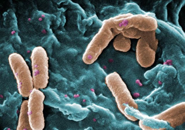
It’s fascinating that Demanèche’s team showed that some bacteria can be encouraged to take up foreign DNA by exposure to artificial lightning, but what about natural populations of soil bacteria? E. coli is not a typical soil microbe (even though some of its relatives are). Can electroporation occur in actual soil bacterial communities? Hélène Cérémonie and her coauthors [3] subsequently isolated bacteria from natural soil. Then, using a similar apparatus as Demanèche et al. (2001), they found that several strains of these bacteria (belonging mainly to the genus Pseudomonas) were also capable of lightning-induced transformation.

An example of a species in the bacterial genus Pseudomonas (P. aeruginosa). Image source: Centers for Disease Control and Prevention’s Public Health Image Library (Wikimedia Commons)

Of particular interest, the bacteria in this latter experiment were most receptive to lightning-induced DNA uptake while in their stationary growth phase. Bacterial cultues in the lab usually exhibit a J-shaped growth curve (when we plot population versus time), caused by the cells dividing slowly at first (as they adapt to their new environment, often requiring the synthesis of certain enzymes to make use of the food with which they are provided), then speeding up their reproduction when they are maximally ready to use their resources, and finally slowing down again as their resources run out. When their food runs out, their population reaches a plateau (their growth is stationary), and they begin to die as wastes accumulate in their environment. Many bacteria respond to limited resources by halting most of their cellular functions in order to survive bouts of hardship. It is in this kind of dormant state that soil bacteria are often found in nature; ready to take advantage of bursts of resources between long lean times.

It is interesting that the potentially evolution-accelerating phenomenon of lightning-mediated transformation was observed to be most effective in the state in which bacteria are commonly found in nature. It is in this stationary phase that bacteria should derive the greatest benefit from the acquisition of a new physiological ability.

The Bottom Line: Implications for Evolution

What exactly does lightning-mediated transformation imply for evolution of life on earth? It means that a widespread natural power source is available to fuel a process that can accelerate the rate of evolution over what we would expect if life forms did not trade ‘genetic ideas’ by HGT (recall, horizontal gene transfer).

Because of HGT, the whole world (or at least large portions of it) can be seen as an interconnected laboratory where advantageous mutations may be sampled and combined with other advantageous mutations in unrelated species, giving rise to unexpected and otherwise highly unlikely combinations of adaptations and advancements. Lightning may further speed this up.

Without HGT, each independent lineage of organisms would need to ‘invent’ by evolution all of their capabilities to, say, utilize various food and energy sources, that they acquired since their last shared common ancestor. The swapping of genes that constitutes HGT drastically reduces the need for a high incidence of beneficial mutations. In principle, a new beneficial trait need only evolve once in a single species and be shared by many species via HGT.

The other reason that HGT can accelerate evolution is that not all advantageous mutations need be lost upon the death of the microorganism in which they arise, but can be resurrected if the relevant genes are acquired from the remains of the dead microorganism by a living microbe in its vicinity upon exposure to electricity in the soil (as one mechanism of acquisition). This is akin to the discovery and publication of a trove of lost unpublished manuscripts of a great novelst after the novelist’s death.

Thus far, very little research appears to have been done on lightning-mediated transformation, this naturally occurring form of electroporation. It will be interesting to watch whether further research can give us a better understanding of how much and under what conditions this unusual electrical phenomenon is involved in enhancing HGT, and by extension, the rate of evolution in the natural world.

References

1. Neumann E, Schaefer-Ridder M, Wang Y, Hofschneider PH (1982). Gene transfer into mouse lyoma cells by electroporation in high electric fields. EMBO Journal. 1(7): 841–845. (FREE DOWNLOAD)

2. Demanèche S, Bertolla F, Buret F, Nalin R, Sailland A, Auriol P, Vogel TM, Simonet P. (2001). Laboratory-Scale Evidence for Lightning-Mediated Gene Transfer in Soil. Applied and Environmental Microbiology. 67(8): 3440-3444. (FREE DOWNLOAD)

3. Cérémonie H, Buret F, Simonet P, Vogel TM. (2004). Isolation of lightning-competent soil bacteria. Applied and Environmental Microbiology. 70(10): 6342-6346. (FREE DOWNLOAD)

Posted in Uncategorized | Leave a comment

Fleshing Out the Past: HMNS Hall of Paleontology

There’s a reason that it’s been a long time since the last Evolutionary Routes post. When I’m not writing about the results of funky scientific research, my artistic alter-ego takes on scientific illustration projects with museums and book publishers. In fact, these days, scientific illustration (especially a subset focusing on prehistoric subjects and coined ‘paleoart’ by paleoartist Mark Hallett) makes up the bulk of my work.

For the last several months, I’ve been collaborating with the Houston Museum of Natural Science in Texas to roll out their enormous new Hall of Paleontology, which is set to be one of the top five paleontology exhibitions in the United States. It opened on June 2 amidst near record-breaking Houston temperatures. At around 36,000 square feet, it’s bigger than a football field, more than 50 feet tall and packed with over 30 dinosaur skeletons, plus hundreds of smaller critters, all mounted in dynamic poses apparently ready to pluck unwary visitors from the labyrinth of hall below.

The skeletons, trace fossils, text and murals chronicle the evolution of life on earth from the Precambrian to the Holocene. My part in the project was to create fifteen colorful back-lit murals, each as tall as a person, to flesh out the prehistoric creatures and their environments, helping visitors to visualize what these other-worldly living things looked like in ages long gone. This exhibit is definitely a must-see, and the short safari on which I’ll take you here only highlights an aspect of one facet of the exhibition, namely the development of the murals.

Yours truly at the opening of the new Hall of Paleontology at the Houston Museum of Natural Science, performing the rarely photographed “hand waving” behavior that is characteristic of scientists when laying out the details of a topic about the lack of which they know an immense amount — in this case, the ecology of the Permian in Texas.

In a museum commission like this, a lot of effort is put into ensuring as great scientific accuracy as possible. This is where the dialogue with experts begins, well in advance of the first rough sketch. The interaction takes two forms: studying published scientific research and dealing directly with curators. There are reams of fascinating research papers on several ancient ecosystems and the biological elements that constitute them, taking out a lot of the guesswork about how these environments would have felt to walk through. However, many fossil ecosystems are relatively new to science. In these cases, it is helpful to gain additional insight and fill in some of the gaps of knowledge by talking directly with the scientists who are involved in doing the first-hand work to uncover the ancient past.

Devonian mural. A pair of Dunkleosteus spar while Acanthostega takes cover midst the Archaeopteris debris.

I love working with museums to endeavor to create works of art with high scientific fidelity. The diversity of people with whom I get to interact is very great, and there are some highly memorable characters. Possibly the personalities that fall at the farthest extremities of the normal curve are the scientists. We’re an eccentric lot, and paleontologists  are no exception. Torrents of enthusiasm can bubble to the surface when they describe the worlds that their work reanimates in their minds.

Permian mural (1 of 2). Sail-backed synapsids such as Dimetrodon feast on the shark Xenacanthus, while more basal tetrapods such as Eryops hunt boomerang-headed Diplocaulus in ephemeral streams also inhabited by Trimerorhachis.

As an artist, it’s very entertaining and inspiring to talk with people such as Dr. Bob Bakker (Curator of Paleontology, and one of the main scientific minds behind the exhibit) when they elucidate, say, the likely goings-on of a typical day in Permian Texas. I always feel motivated to illustrate after such discussions. Even on the phone, I can practically hear him gesticulating enthusiastically as he describes giant sail-backed predators snacking on sharks in crowded streams.

Permian mural (2 of 2). A pair of wolf-like gorgonopsids called Inostrancevia scuffle over the carcass of a dicynodont.

And it’s not just words that are exchanged. Anyone who’s read his works such as “The Dinosaur Heresies” knows that Dr. Bakker is also an accomplished paleoartist. He provided numerous concept sketches for the exhibit, establishing the intended postures in which the animals’ skeletons were to be mounted. (It’s no coincidence that many of the skeletons on display remind one of his drawings.) I often based the postures of the species in the murals on such sketches. One of the best examples is the Dimetrodon/Xenacanthus reciprocal attack tableau in the Permian mural.

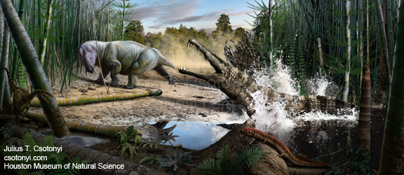
Triassic mural (1 of 2). The archosaur Smilosuchus lunges from the water in an effort to snag the dicynodont Placerias for breakfast.

Once everybody on board is happy with the planned content of a scene, I generate the first rough draft of a mural. At this point, a good dialogue between artist and experts is vital, because the first draft usually requires a lot of revision until it’s given the nod of approval. Everything from the relative position of animals to the thickness of a snout based on new fossil evidence can require alteration of the rough draft, and usually several rounds of review and revision take place.

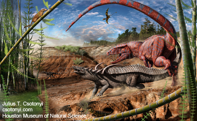
Triassic mural (2 of 2). A hungry rauisuchian, Postosuchus, attempts to sidestep the spiny armour of its intended meal, the bizarre herbivorous crocodile-related aetosaur known as Desmatosuchus.

Most revisions simply increase the accuracy of the images. However, there are other factors to consider, some of which you just don’t see coming. In what is by far the most unusual example, the Triassic scene depicting a hungry Postosuchus attacking an armoured Desmatosuchus was based on a dynamic concept sketch by Dr. Bakker in which the former steps over the tail of the latter during the scuffle. My rough draft of the mural rotated the scene so that the viewer sees the action from behind and below, allowing for some interesting compositional effects. However, my draft was just slightly ‘off’ in the animals’ mutual proximity, and an unintended side-effect of the particular angle chosen was that the animals could potentially be interpretted as engaging in amourous rather than predatory behaviour; by offsetting them slightly from each other (it was hoped), potential misunderstandings would be averted.

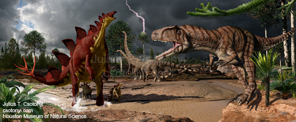
Terrestrial Jurassic mural. In this Morrison formation scene, a protective mother Stegosaurus provides four good spiny reasons for an Allosaurus to move along while a pair of Diplodocus browse in the background.

Once the revised rough draft is given a unanimous thumbs-up, I proceed to rendering the details in high quality. Although I frequently cater to clients’ requests to provide work that has a specifically ‘painted’ feel (watercolor, acrylic, etc., such as my  Cretaceous murals in the new Dinosaur Hall at the Los Angeles County Museum of Natural History), my preferred artistic style is characterized as hyper-photorealistic. I achieve this by harmonizing a variety of digital techniques, from digital painting to photographic compositing, as in the movie industry. The latter requires me to acquire photographs of the geological, floral and faunal elements for the images, not only for visual reference but also incorporation of image content.

Marine Jurassic mural. Amidst a hanging garden of the crinoid Seirocrinus (one interpretation of the configuration of such fossils), a Stenopterygius mother is orbitted by six of her young as she dispatches a Harpoceras, while the marine crocodile Steneosaurus eyes a lone young ichthyosaur that has wandered off to investigate another ammonite.

But I can’t just snap a photo of a grassy field, smack a Triassic animal into it, and grin with satisfaction. A scientific illustrator must strive not only to portray anatomically accurate animals, but also geologically appropriate associated paleobotanical communities. Plants matter as much as animals, despite the tendancy of some artists in the past to focus almost exclusively on the animals, either placing them in a “parking lot” devoid of plants, or depicting anachronistic plant communities (such as grass in the Triassic).

Terrestrial Cretaceous mural. A species rich image that spans a time from the Campanian (right) to the Maastrichtian (left) in North America, this mural depicts several tableaus of activity, including a confrontation between a Tyrannosaurus and the wonderfully preserved mummified Triceratops known as “Lane” (center). The integument of Lane’s back is a mosaic of large polygonal scales, interspersed with giant plate-like scales that possess raised knobs in the center, some of which appear to be the broken bases of structures that may have been longer projections. Discussions with the paleontologists involved in the project led to the depiction of this Triceratops with bristles emerging from these enlarged scales. This interpretation is speculative, but the presence of long quill-like or filamentous structures is precedented in the skin of some other ornithischian dinosaurs, such as the ceratopsian Psittacosaurus and the heterodontosaurid Tianyulong.

Attention to this kind of detail while maintaining photorealism requires me to travel to places on earth that host what are known as analog communities, whose overall structure and species composition closely resemble certain prehistoric communities. For example, the Taxodium (swamp cypress) communities depicted in the Maastrictian (latest Cretaceous) half of the terrestrial Cretaceous mural come from photos of swampy or slow-flowing river locations in eastern Texas and South Carolina. Some ecosystems from the Cretaceous looked a lot like certain subtropical or temperate forests and wetlands of today. Such landscapes may only require small adjustments to agree with scientists best models of the compostion of prehistoric ecosystems.

Marine Cretaceous Mural. The Western Interior Seaway teems with reptilian life, including the giant marie turtle Archelon, which nips at a flipper of the giant mosasaur, Tylosaurus, a colossal relative of the living komodo dragon.

However, one of the neatest things about prehistoric biological communities brought about by evolution is that the farther back in time you look (i.e. the deeper in the strata of the geological column), the less familiar and more alien the landscapes look. This is not only because the composition of species differed, but also because the representatives of familiar goups of plants looked very different from those of today. There has been a lot more time for evolution to mold the appearance of these organisms since their deepest ancestors. Therefore, in many cases, I cannot simply photograph an existing landscape and only slightly modify this analog to make it agree visually with what we expect to have seen millions of years ago in the living version of the fossilized ecosystem. When the very plants themselves have no visually similar living representatives, I must build up these species from scratch, and then incorporate the elements of my work in an appropriately arranged community. Some mural landscapes are therefore much more a mosaic of work than are others. The Triassic and Permian murals are good examples of this process. Notice the segmented Neocalamites and Equisetites trees. They’re relatives of today’s horsetail plants, but they grew to around 20 feet (6 m) tall, and looked considerably different from modern horsetails.

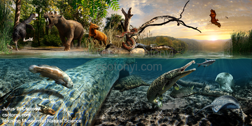
Eocene mural. The Green River formation hosts a plethora of early mammals (such as the weird sabre-toothed Uintatherium) and other animals that shared their lakeshore environment, including the large armoured gar Atractosteus, the freshwater ray Heliobatis, the giant ‘terror-bird’ Diatryma, an early snake known as Boavus and one of the earliest bats, Icaronycteris.

Even the appearance of the nonliving parts of the landscape must be tailored, for paleontologists and geologists can deduce from the chemical composition and physical structure of the matrix material containing fossils what the ground and landscape were like at the time that the sediments were deposited. For example, sandstones sometimes indicate dunes whereas shales generally result from muddy surfaces. The matrix and state of preservation of fossils within it can even tell us about the climate that predominated when the creatures died.

Oligocene mural. The White River formation preserves a strange ecosystem in which giant carnivorous relatives of pigs, known as entelodonts (here represented by Archaeotherium, center) probably chased down diminutive ancestors of familiar modern mammal groups, such as the early horse Mesohippus.

Physical and chemical cues within the rocks (as well as the species compositon of plants and animals whose fossils exhibit adaptations to certain environmental conditions) tell paleontologists that, for example, the Permian beds of Texas that yielded the Dimetrodon nicknamed “Willie” were deposited in a region that experienced seasonal peaks in precipitation. These monsoon-like rains periodically generated streams in which feeding frenzies appear to have taken place during the emergence of some animals from drought-evading burrows in the mud (just as lungfish do today). Such a ‘breakfast bonanza’ is depicted in the Permian mural, where sail-backed synapsids (relatives of early precursors of mammals) devour bizarre spined eel-like freshwater sharks known as Xenacanthus.

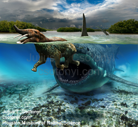
Miocene mural (1 of 2). The world’s largest known shark, Carcharodon megalodon, stalks an early proboscidean, the ‘shovel-tusker’ known as Platybelodon, in the shallows of a lagoon. The gape of the shark is 11 feet (3.3 m) wide.

At the risk of aggravating paleobotanists, the illustration of animals often receives special attention. Some of what drives this faunal focus is that charismatic animal fossils are often placed at the center of displays, and murals tend to highlight these stars of the show. Patrons are generally more excited by animal fossils than those of plants. This is like the phenomenon of children peering into terrariums in search of animal life (especially active animals) and looking straight past the fascinating plants. In some ways, it’s too bad that so many of us seem to take a much greater interest in fauna than flora, because we thus miss out on a tremendous number of brow-raising tales of botanical natural history. To that end, I would be very interested in illustrating an exhibit that focused primarily on prehistoric plant communities. Still, despite the abundance of faunal fossils, the HMNS team did a very nice job of placing the animals in the proper ecological context, which is a gratifyingly increasing trend in many museums today.

Miocene mural (2 of 2). In this semi-arid steppe landscape of ancient China, one member of a herd of the tusked rhinoceros Chilotherium munches on blooming composites while another harasses a much larger mastodon, Mammut. A pair of the horse Hipparion protest the disturbance.

To flesh out the animals in the murals, I relied on a number of techniques, from digital painting to sculpting to photographic compositing. When you can’t go out to photograph a 70 million year old Cretaceous mammal, you need to be resourceful to get the job done. In the case of the Didelphodon that appears in the terrestrial Cretaceous mural, I used as a model our Corgi/Jack Russell dog, Wiki (from a Hawaiian word meaning ‘fast’ or ‘quick’, a very apt name for her). In one photoshoot, we persuaded Wiki to engage her ‘begging mode’, which reasonably closely approximates the clam-clutching bipedal stance of one of the Didephodon skeletal mounts in the exhibit. By working from these photographs as visual reference for the way that lighting falls on different parts of a small furry animal while relying on the Didelphodon skeleton as guide, I was able to build up an anatomically and posturally accurate reconstruction of Didelphodon in a photorealistic manner.

Pliocene/Pleistocene mural (1 of 2). One of a pair of murals depicting the great American interchange of mammals, in which many species migrated between North and South America to establish more modern species distributions between the continents when the land bridge formed. Here, a Xenosmilus leaps out of the way of the armoured club-like tail of the giant armadillo relative Glyptodon, while the aptly named bird, Titanis, lopes off to the left.

To be sure, a lot of pressure can build up as the deadline for submission of artwork nears and when it still feels like I am months away from completion. Inevitably, the candle begins to burn not only at both ends, but at several points in between. Nevertheless, the best indicator that tells me I’m in the right field of work as a scientific illustrator is that even at the peak of this pressure cooking, I still draw enjoyment from the work of illustration itself. Perhaps it is this gratification from the exciting nature of my job (I get to paint dinosaurs for a living; that never gets old) that helps me reap the energy I need at the home stretch to keep delivering the materials on time, which is crucial to a timely opening.

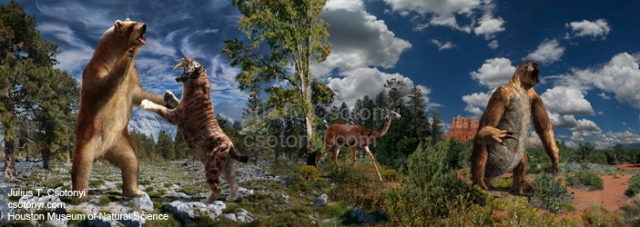
Pliocene/Pleistocene mural (2 of 2). In the second in the American Interchange murals, the huge short-faced bear Arctodus faces off with the sabre-toothed Smilodon, as the gazelle-like camel Hemiauchenia ambles out of the way. The giant ground sloth Eremotherium rises to full height on its hind legs.

The new permanent paleontology wing has grown from a twinkle in the developers’ eyes about five years ago, to a reality today. It was a pleasure to work with the paleontologists and exhibit designers to create the colorful visual elements of these murals, which depict about 90 species of prehistoric animals all told. The HMNS Hall of Paleontology is a permanent exhibit, viewable at the Houston Museum of Natural Science in Hermann Park, located in central Houston, Texas.

Featuring more of my murals, another exhibition, “Ultimate Dinosaurs: Giants from Gondwana” opens later this month (June 23, 2012) at the Royal Ontario Museum in Toronto, Canada for a limited time.

Posted in Uncategorized | 9 Comments

Fish Out of Water

The highly productive sciences of biology and paleontology are rapidly bridging the gaps in our knowledge about how disparate life forms diversified to fill all manner of niches on earth. Both in the fossil record and among living biological diversity, biologists and paleontologists are providing myriad colorful examples of ‘transitional’ life forms on a nearly daily basis. These researchers are making life increasingly difficult for bloggers of evolutionary biology like myself. Just how are we supposed to satisfy our obsession of discussing all these finds and still have time in our schedules? Well, now that I’ve ranted, on to the news:

A Fish With Grasshopper Dreams

Terry J. Ord and Tonia Hsieh just published a delightful natural history study about a fish that is very nearly a fully land animal, for it spends the vast majority of its time out of water, and even actively avoids submersion by waves [1]. It is a marvelous living example of what is often referred to as a ‘transitional form’: a creature that has characteristics that make it evolutionarily intermediate between two well-established groups or life histories (but beware of the limitations of this term, covered at the end of this post). Why is this so significant? To evolutionary biologists, it is no surprise that evolution repeats itself, resulting in multiple concurrent examples of what look like half-finished projects at various stages of ‘completion’. However, proponents of creationism or intelligent design often point to alleged gaps between major groups of organisms (e.g. land-dwelling vertebrates and fish) in an attempt to support the claim that there is no evidence for the evolution of one group from another. Such evidence may have been less abundant in the days when Darwin published his seminal tome, but after 150 years of research effort, this is no longer the case; not by a longshot.

Ord and Hsieh report in detail the behaviour of a perplexing little fish, the Pacific leaping blenny (Alticus arnoldorum), which, as its name suggests, seems to think it’s a grasshopper. Not only does this highly social colony-breeding fish spend more time out of water than submerged, but it escapes capture by rapidly contorting its body like a discharging spring, propelling it several body-lengths away in a single bound (and making capture for study no easy task). Although the skin and gills of this remarkable little fish must still be periodically moistened, for it breathes through them, it is otherwise entirely terrestrial.

Let that thought really sink in. We have here a land-dwelling fish.

Pacific leaping blenny on rocks. (Image: Fastily)

This is a momentous find indeed. However, most people are probably unaware of how many other groups of fish also exhibit adaptations to life out of water to various extents. I review some of these fascinating examples below. Not only do these results catch evolution in the act, providing some of the best examples of living animals that natural selection has moulded into forms transitional between aquatic and land-dwelling creatures. They also lend us a nice view inside the clockwork of evolution, clearly illustrating how it works. Although the first invasion of land by fish during the Devonian Period probably took a different route according to the fossil record, the living examples demonstrate that the transition to land can occur by a number of means. We see several of the ecological factors that can conspire to drive fish onto land.

Flying Fish

Most of us are familiar with one of the most unusual fish to leave water (albeit for very short periods of under a minute): flying fish. These creatures propel themselves with their powerful caudal (tail) fin to keep themselves aloft for over 100 m, and up to 400 m, using their enlarged pectoral fins as air foils as they glide just above the surface of the waves at 70 km/h. This flight mechanism facilitates rapid escape from predators. The much lower resistance of air than water makes the atmosphere a much quicker medium through which to move, requiring much less energy to achieve any given speed than through viscous water.

Flyingfish doing its thing. (Image: NOAA)

Even though flying fish do not reside out of water for long (and certainly not on land), they illustrate one of the driving forces behind the invasion of land by vertebrates: the escape from natural enemies. Their fast motion also facilitates this escape, just like the hopping habit of the Pacific leaping blenny.

Gouramis, Snakeheads, Blennies and Mudskippers: The Precocious Perciformes

The Pacific leaping blenny belongs to a very large successful group of fish, the order called the Perciformes. Many fish popular with anglers belong to this group, including perches. Interestingly, there are several suborders of fish among the Perciformes that display adaptations for survival out of water. One of the common adaptations is the development of lung-like organs, and this is overwhelmingly in response to poor oxygen conditions in their environment that make breathing by gills alone difficult.

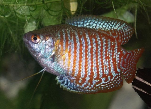
Does a Betta or a kissing gourami inhabit your aquarium? If so, then you have an air-breathing fish. Members of the suborder Anabantoidei, including popular aquarium fish such as gourami and Siamese fighting fish, are known as labyrinth fish. The name refers to an interesting adaptation that the group shares. They possess a highly branched and folded extension of the gills, called the labyrinth organ. This structure allows the fish to gulp air at the surface of their often poorly aerated pools to supplement their oxygen intake. The labyrinth organ is richly supplied with blood vessels to soak up oxygen from the gulped air, rather than from water. Water is not good at carrying oxygen as it is, compared to a gas, but stagnant tropical water holds even less. So the presence of a rich supply of oxygen just above the water’s surface provides a strong selection pressure for a creature to develop a way to tap into it. Interestingly, this group of fish has evolved to depend so highly on the labyrinth organ for oxygen that they would drown if prevented from reaching the surface. So yes, you can drown a fish. These fish, anyway.

A type of gourami, a partially air-breathing fish with a labyrinth organ. (Image: Quatermass)

Note that the labyrinth organ is an adaptation to breathing air for the express purpose of circumventing the poor oxygenation of their watery environs, but not for life on land. Thus, here is evidence that an adaptation can evolve for a different purpose that that for which it might ultimately be useful. This is called preadaptation. It illustrates that early land animals may have already been breathing air before making their transition to land. This is a very important observation, and it provides a nice retort to the suggestion that major evolutionary changes (e.g. transition from life in the water to life on land) require too many simultaneous adaptations to occur. In fact, many of these transitions probably took place by such ‘baby steps’.

However, some of the labyrinth fish have another land adaptation up their sleeves. The climbing gouramies (family Anabantidae), as their name suggests can climb out of their pools of water and ‘walk’ short distances to other pools, pushing themselves along with their tails and using their gill plates as support. This is a wonderful adaptation because it allows an aquatic creature to not only escape predators that cannot follow it onto land, but it also facilitates escape from drying pools that would ordinarily doom aquatic species, and also allows them to disperse their genes more quickly and over greater distances than swimming alone would allow. In some species, this ability has made them extremely adept at getting around. The snakeheads (suborder Channoidei: family Channidae) are terribly invasive species in North America, for example [2]. They also possess a labyrinth organ, allowing them to stay alive breathing air for up to four days. In this time, they can cross moist ground for distances up to 1/4 mile.

Snakehead. (Image: Virginia Department of Natural Resources)

More familiar to most people are mudskippers, bizarre-looking members of the gobies (suborder Gobioidei), a sister group to the blennies. Mudskippers look for all the world like amphibians, with their raised eyestalks and their habit of sitting on rocks and branches above the water line, with only their tails soaking in the water in order to keep their skin moist. Like the Pacific leaping blenny, they are also capable of catapulting themselves great distances (up to 60 cm or 2 feet) into the air. These weird animals breathe air by a number of means: (1) through the moist skin, enriched in fine blood vessels called capillaries, (2) through an especially capillary-rich area in the buccopharyngeal cavity (a region at the back of the mouth and throat), and (3) through their specially adapted gill chambers, which are sealed shut to retain water when the fish moves onto land [3]. The air is swirled around in the gill chamber by rotation of the large eyes, which are used as pumps. Mudskippers also show a wonderful example of how the pectoral fins can be used for locomotion on land. The special elbow-like bends in the fins allow them to be used to prop up the animal and propel it forward.

Mudskipper. (Image: Quartl)

Electric Eels, Lungfish, Walking Catfish, Bowfins, Eels

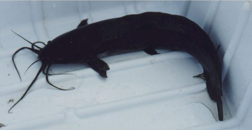
Some manner of terrestrial adaptations have arisen in nearly 20 diverse groups of modern fish, including ‘walking’ catfish and eels that can move between bodies of water, breathing air during the journey.

Walking catfish. (Image: USGS)

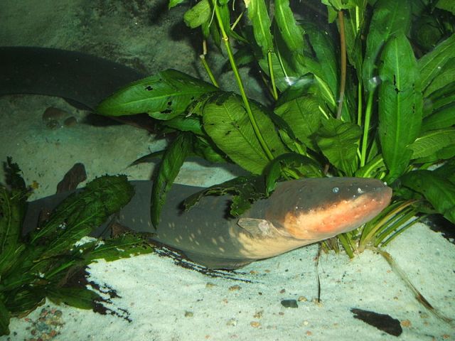
Everyone knows what gives electric eels (Electrophorus electricus) their name — they can deliver 500 watts of electric energy to stun their prey, a lethal dose for humans. But how many of us know that electric eels are obligate air breathers, needing to surface every 10 minutes or so to gulp air for oxygen exchange in an oral cavity [4]. They get about 80% of their oxygen this way!

Electric eel. (Image: Steven G. Johnson)

The air breathing organs of an ancient group of fish called bowfins (order Amiiformes) are derived form the swim bladder, which is a special organ used by bony fishes to maintain neutral buoyancy (float) in the water. In the bowfin, the swim bladder is lined with capillaries and used to help oxygenate their blood because they live in slow moving and oxygen-poor waters. The significance of this adaptation is that the air bladder is homologous with our lungs. In other words, it derives from the same tissues during development, and our lungs are probably evolutionarily derived from swim bladders of early fish.

Bowfin. (Image: Stan Shebs)

Lungfish (subclass Dipnoi) take this vascularization (lining with capillaries) of the swim bladder to an even greater extent. Their swim bladder is divided into a great number of smaller sacs, thus considerably increasing the surface area for gas exchange. As their name implies, these structures serve as lungs. Their lungs are an adaptation to their highly marginal or seasonally harsh environment. The lungs help them to survive for months at a time through the dry season during which their bodies of water dry up. During this time, lungfish breathe only air, while they remain entombed in cocoons in the mud with only a breathing tube connecting them to the atmosphere.

Marbled lungfish. (Image: Opencage)

The Fossil Record: Tiktaalik, Ventastega and Friends

This post deals mainly with living examples of evolutionary intermediates between aquatic and terrestrial species, and I will cover the fossil record in more detail in a future post (and I have also briefly discussed the fascinating story of the discovery of Tiktaalik, a wonderful example of an intermediate form between fish and amphibians, in the post “Wasp-Infested Dinosaur Eggs and the Science of Paleontology“). Even better examples are still being found, bridging the gap between even Tiktaalik and amphibians. One example is Ventastega, which has shoulder and hip characteristics that place it between Tiktaalik and early amphibians such as Acanthostega [5].

“God of the Gaps” Meets Achilles and the Tortoise

Perplexingly, despite the accumulating finds of transitional forms that help patch holes in our knowledge of the fossil record, proponents of creationism sometimes view this development as simply an increase in the number of gaps that need to be filled; each gap is simply bisected by a newly discovered transitional form, making two holes out of one and multiplying the burden of proof for scientists! This absurd conclusion is a spin-off of the theological viewpoint popularly referred to as the “God of the Gaps” argument. It is of course a simple logical fallacy, akin to the Achilles versus tortoise paradox, in which the ancient greek runner Achilles appears to be unable to catch up to a tortoise with a head start, but only if the progress of Achilles’ is dissected into incrementally shorter segments delineated by the advance that the tortoise’s makes each time that Achilles catches up to the point occupied by the tortoise on the previous iteration. To escape from the paradox, we must realize that the problem lies in believing that the later shorter segments of advance were given the same temporal importance as the earlier longer ones. Similarly, it is crucial to realize that the closing of the evolutionary distance between established life forms is more important than the number of the gaps between them, because a shorter gap in fact presents a proportionately smaller problem.

Black (and Numerous Shades of Grey) and White

Another complaint that evolutionary biologists face regarding transitional forms is based on the erroneous view that the world is only black and white, that any given transitional form must be viewed as either belonging to one group or another, and that there is no intermediate grey area that any organism can occupy. This view is probably most vocally expressed in discussions of evolutionary anthropology, in which creationists that refuse to accept the evolutionary descent of humans from earlier primates attempt to mask the clearly quantifiable gradient in characteristics between humans and ancestral primates by choosing to classify a hominid fossil as either human or non-human ape (more on this in a later post).

Taxonomy (the science of categorizing and naming creatures) may sometimes appear to try to force a species into one group or another. However, the purpose of taxonomy is mainly to allow us to distinguish species from each other by a list of carefully measured characteristics. However, taxonomy was established as a field of study before the evolutionary relationship between organisms was discovered, and therefore taxonomy lacks a rigorous quantitative estimate of how closely related two groups are to each other. Under the encompassing field of systematics, the hierarchical classification system of taxonomy must strive to maximally reflect the results of a sister branch of study called phylogeny, which attempts to delineate the ancestor/descendent connectivity of different life forms by measuring both the topology (branching pattern) and evolutionary distance between these groups. Phylogenetic and evolutionary studies that show a clear gradual progression in the development of a trait over time (either in the fossil record or in rapidly evolving living systems) highlight a problem that taxonomy by itself is hard-pressed to resolve: it becomes ever more difficult to pigeon-hole a life form into one of two groups if it is evolutionarily intermediate between the two. Taxonomy may be up to the task, but it becomes increasingly impractical as we near the midway point, for the number of characteristics that need to be clearly defined to differentiate the two groups rises, and at some point, arbitrary decisions need to be made regarding, say, the length of a bone in assigning it to one or the other group. Ultimately we can maximally resolve the evolutionary lineage to the point of individuals in the line of descent. Those are the quanta. But how do we know at which individual (had we all of the fossils in our hands even!) to decide to draw the line between fish and amphibians?

This problem is well illustrated by trying to decide at what point a red-to-blue gradient of paint becomes unambiguously blue. Even if we decided to define the border by a particular exact wavelength of the light, our staunch position does not eliminate the fact that the red and blue ends of the gradient are, in fact, connected by a smooth gradient, not a sharp discontinuity. Such is the case with the fossil record. Even if one decides to call one fossil a fish and another an amphibian, it is a misleading distinction if it does not consider the fact that there is a gradual progression of forms that are most parsimoniously explained as expressing gradual evolutionary change.

How Meaningful is the Term “Transitional Form”?

It’s important to add a healthy grain of salt to our discussion of transitional forms. It is reasonable to view every species, especially those from the fossil record, as being transitional; it is statistically unlikely that any given fossil represents the last individual or species in its given line of descent before that particular branch went extinct (and most branches do). The process of fossilization is so sporadic that we are not likely to predictably come across something that is this precisely positioned in the line of descent of a group — except perhaps at a well-defined boundary demarcating a mass extinction beyond which a group is highly unlikely to have survived, (but even some of these boundaries may not have been guillotine sharp with respect to extinction, e.g. some dinosaurs may have survived the asteroid impact 65.5 million years ago by as much as 700,000 years, which I discussed in a previous post, “Dating Dinosaurs“). The only creatures that we can hold in our hands and be certain to be currently at the true terminus of their own lines of descent are living specimens.

But although the vast majority of fossilized organisms are almost certainly transitional between ghosts of evolution that fossilization never preserved, perhaps none of them are truly transitional in a geneological sense between two specific fossils that we can hold in our hands which nonetheless look like the two states of evolution that our specimen bridges. As pointed out in a previous post (“Archaeopteryx: Should Feathers Fly Over Its Fall?“), any given fossil specimen is most likely a member of a short evolutionary branch from the main line of descent in which we’re interested, because it is statistically unlikely in the extreme that the individual preserved for millions of years belonged to the infinitesimally narrow bridge of descent connecting one ancestor with another distant descendent. This situation is a natural result of the incredible bushiness of the tree of life; there are far more terminal twigs than main connective branches.

So, should we even use the term “transitional form” if it is so ambiguous? I have chosen to employ it in this post mainly because it is a familiar term, but I have endeavored to explain its limitations. Nevertheless, semantics aside, the evolutionary intermediacy of organisms between highly different forms and lifestyles is very real, and both the fossil record and the census of living organisms today provide wonderful examples to support this conclusion.

References

1. Terry J. Ord and S. Tonia Hsieh. (2011) A Highly Social, Land-Dwelling Fish Defends Territories in a Constantly Fluctuating EnvironmentEthology, 26 DOI:10.1111/j.1439-0310.2011.01949.x

2. Böhme, Madelaine (May 2004). Migration history of air-breathing fishes reveals Neogene atmospheric circulation patternsGeology 32 (5): 393–396. doi:10.1130/G20316.1

3. A. Graham JB, ed (1997). Air–breathing Fishes. Evolution, Diversity and Adaptation. San Diego California: Academic Press.

4. Johansen, Kjell (1968). Gas Exchange and Control of Breathing in the Electric Eel, Electrophorus electricusZ. Vergl. Physiologie (Springer Berlin / Heidelberg) (Volume 61, Number 2 / June, 1968): 137–163.

5. Per E. Ahlberg, Jennifer A. Clack, Ervins Luksevics, Henning Blom, Ivars Zupins. (2008) Ventastega curonica and the origin of tetrapod morphologyNature 453 (7199): 1199 DOI: 10.1038/nature06991

Posted in Uncategorized | 2 Comments

The Taming of the Chloroplast

A fascinating study [1] has recently underscored the hypothesis that our mitochondria, the power packs of our cells, share an ancient kinship with a particularly nasty disease, typhus. Put plainly, the same bacterium may have been the granddaddy of each. This result surprisingly implies that we’re all partly bacterial, and not only in a genetic sense but also structurally. In the last post (“Frankenstein’s Batteries“), I covered the story of how a forced cohabitation with our one-celled ancestors could have made the originally disease-causing bacterium change its stripes and give rise to the essential mitochondria that now power our cells. Dedicated components of our cells (organelles) have been acquired via the process of endosymbiosis, in which one organism lives within the body of another in order to gain access to benefits such as a reliable source of nutrients or protection from the elements.

But many people, especially if they have been courted by the ideology of creationism or intelligent design, might respond: “Fascinating story, but too bad you don’t have evidence of intermediate stages of the process. All you have are fully parasitic microbes and fully integrated organelles, right? Can you point to any living candidates of in-between stages?” To this question, an over-enthusiastic biologist — not unlike myself — would bristle with excitement and answer: “Glad you asked! Let’s rectify this problem and fill the alleged gaps with some colorful living examples.”

So let’s look at some weird and wonderful examples of living intermediate stages in the taming of another organelle, the chloroplast. But first, let’s consider how chloroplasts appear to have evolved, and the evidence for their evolutionary origin.

Cells of the moss Plagiomnium affine, showing the numerous brilliant green disk-shaped chloroplasts inside each cell. (Image: Kristian Peters -- Fabelfroh)

Plants and Algae: Hunters-Turned-Farmers

Plant and algae cells have mitochondria, the same as we do. (Recall, from the last post, that mitochondria are the power plants in our cells, churning out useful energy by ‘burning’ the food molecules that we ingest.) But the ancestors of plants had an even better idea than did our animal cells’ ancestors. (Of course, you need to read “idea” in a figurative sense only — cells have no conscious volition or thought processes.) Plants’ ancestors acquired an improved version of an energy generating device — a chloroplast — by engulfing cyanobacteria, a photosynthetic group of bacteria that are also known as “blue-green algae”. The chloroplast differs from the mitochondrion in that it appears to have arisen not from the invasion of a host cell by a parasite, but from a predator playing with its food before digesting it. Nevertheless, it’s a splendid example of endosymbiosis.

In those early days, plant cells’ ancestors did not possess the green color that they do today. Whatever green hues they did sport probably came from the cyanobacteria that they ate, and it lasted for the short time before they proceeded to digest the cells, pigments and all. Cyanobacteria are good sources of food because they are full of chemical energy. Cyanobacteria use the green pigment chlorophyll (in collaboration with red, orange and yellow pigments called carotenoids) to convert, or transduce, solar energy from the sun into useful chemical energy. They do this by cocking the hammer, as it were, on high-powered molecules such as adenosine triphosphate, or ATP. But they also store some of the energy from light in the energetic chemical bonds within sugar molecules. This way, they not only get abundant free energy from the sun, but are also able to shelf some of it in a stable form that their mitochondria convert into ATP later at night, when the sun is not around. Their solar-powered habit made cyanobacteria palatable to other types of cells in their environment — predatory cells. These predators feasted on cyanobacteria for countless millions of years. But then a revolutionary event occurred. It may have gone something like this:

A predatory eukaryotic cell slurped up cyanobacteria from the warm surface of a pond, lake or ocean, engulfing them by phagocytosis — i.e. wrapping a membrane around them and then pumping digestive enzymes into the membrane to dismantle the cyanobacteria. The simple digested molecules were then fed into mitochondria, which turned them into energy (ATP) for the cell. But the cyanobacteria continued to make sugar by photosynthesis for a short time before they were digested in the phagocytosis membrane. Some of this sugar leaked out into the surrounding cell, and for a short time before digestion, the predatory cell enjoyed photosynthetically-manufactured food, courtesy of the cyanobacterium’s last gasps. Some cells refrained from digesting their captured cyanobacteria because it turns out that if they stockpiled a whole lot of these membrane-coated cyanobacterial ‘candies’ and let them continue to live while restraining their rate of reproduction, then the predators (now hosts) could soak up so much sugar from their cyanobacterial captives that they didn’t even need to expend energy to ingest more cyanobacteria. They could just bake in the sun all day long and let their cyanobacterial vending machines pump out sugar for their mitochondria to convert into energy.

And so the chloroplast was born, a membrane-enclosed organelle in plant and algal cells that carries out photosynthesis. Of course, cells did not make any conscious decisions to refrain from digesting cyanobacteria. But when several ways of life are available to an organism, the more economically prudent option (with a high ratio of benefit to cost) results in more offspring, making this way of life ever more prevalent in the population if it can be inherited genetically. Retention of living cyanobacteria offered the great opportunity to abandon the expensive business of hunting at the small cost of foregoing the energy from digesting the whole cyanobacterial cell. It was a little like the transition that our own human ancestors made thousands of years ago: farming presented a good return on the investment of giving up hunting.

Cross-section of a generic chloroplast, the organelle in which photosynthesis occurs in plants and algae. Its structure and genome is very similar to that of ancestral cyanobacteria. (Image: Miguelsierra)

Evidence for Cyanobacterial Ancestry of Chloroplasts

As for mitochondria, there’s evidence of a bacterial origin for chloroplasts. Like mitochondria, chloroplasts possess their own DNA (called the plastome), which is a circular loop, more like that in bacteria than in the eukaryotic host cell. This DNA is severely reduced, containing only about 5% as many genes as free-living cyanobacteria, but the genes that are present are similar to those of their cyanobacterial ancestors. And the missing DNA? Amazingly, much of it has been transferred from the chloroplast to the host’s own chromosomes in the nucleus. This extensive genetic cross-wiring illustrates that stable endosymbiosis has caused an impressive amount of the labour of living to be divided between the host and its photosynthetic guest.

Farming photosynthetic organisms by engulfing them and then holding them captive to soak up their sugary cell products is such a good strategy for survival that there is evidence that it has taken place multiple times in the past in different groups of cells. Molecular biologists can recognize at least two genetic lines of chloroplasts, meaning that cyanobacteria were engulfed and successfully ‘domesticated’ on at least two separate occasions (in the Archaeoplastida, which includes plants and algae on the one hand, and in the amoeboid cell Paulinella on the other hand). Furthermore, within the main Archaeoplastida genetic line, we see evidence of not only primary endosymbiosis (establishing chloroplasts by engulfing naked cyanobacteria), but also secondary endosymbiosis. This means that some algae, such as Vaucheria, appear to have engulfed an algae that had in turn engulfed a cyanobacterium ages ago. The whole arrangement now resembles Russian nesting dolls, with the original cyanobacterial descendent wrapped in four concentric membranes corresponding to the two membranes of the chloroplast nested within the cell membrane of its primary host and wrapped in the feeding membrane of its secondary host. In some cases (e.g. cryptomonads), the original nucleus of the engulfed algal cell is still found squashed between the membranes of the chloroplast.

An evolutionary tree showing the relatedness of different lineages of chloroplasts in living groups of organisms. Generally, each major lineage originates from a unique event of endosymbiosis, the establishment of a stable association between a host and either a cyanobacterium or an algae that already harbours a chloroplast. (Image: Nova)

Almost a Chloroplast: Intermediate Stages in Evolution

But there’s a catch: if this predator-gone-plant way of life was so attractive that it arose more than once, then we should expect to see it happening today as well. We should see some intermediate stage of chloroplast taming going on. Evolution doesn’t normally just work once and then stop. Predatory cells that feed on photosynthetic cells should still be embarking on similar collaborative business ventures with their food. So where are they? They’re all around us, it turns out.

A Fungus Took a ‘Lichen’ to Some Algae…

Lichens are a weird chimaeric life form with an almost absurdly robust ability to survive under extreme conditions, such as the soil-free surface of wind-lashed rocks on the peaks of mountains, tomb stones,  tree trunks, old rusting automobiles, soil, sand, etc. They derive their versatility partly from their compound nature: lichens consist of a durable fungus (normally a heterotroph, which gets its food from digesting other organisms) whose internal cells are modified into finger-like formations that clasp single cells of cyanobacteria or green algae (like a long-fingered basketball player holding a ball between dribbles), and forcing the membranes of the algae to leak out some of the sugars that it produces. The lichen is thus effectively a photosynthetic fungus, powered by the sun.

A lichen called Xanthoparmelia growing on basalt, a volcanic rock, on O'ahu, Hawaii. The shape of the creature is determined by the fungus, and the subtle green color is due to algae held captive under the upper cortex (skin-like layer) of the fungus. The color is only faintly green because light does not penetrate as easily through the lichen's cortex as it does through the cells of a plant leaf, so most of the color is due to the fungal portion of the creature. Lichens are more obviously green when they are wet, partly because their tissues are more transmissive to light under those conditions. (Image: Eric Guinther)

Lichens are interesting in that the algae do not grow inside the fungus cells (unlike a plant, which has cell-bound chloroplasts), but are held within the body of the many-celled fungus nonetheless. The relatively primitive nature of the association is also underscored by the fact that the algal or cyanobacterial cells have to be captured from the environment each time that the fungus portion reproduces from spores. However, it may be hard to find the algae that it needs to survive in some of the wretched environments in which it lives, and the lichen may die out if it needed to catch its food-producing algae each time it reproduced. So the lichen solves the problem by also being able to reproduce by fragmentation, and by releasing little balls of fungal cells that are already clasping algal cells.

Corals and Anemones: Alternative Green Energy on the Reef

Animals are usually pretty good at getting around and hunting for their food. But say you’re an animal without legs or a brain — not unlike a coral or sea anemone in the ocean. This predicament makes hunting for your food pretty difficult, and you’re forced to rely on laying in wait for snacks to come bumbling along and to get close enough for you to ensnare them in your tentacles. So you’d better hope (also a trifle difficult without a brain) that you live in a heavily populated lagoon, with lots of “accidental” feeding opportunities. It wouldn’t be a bad idea to have a backup plan in case your neighborhood experienced a drop in property value and stopped attracting tourists.

Two examples of mutualism, an interaction between species that mutually benefits each. In the first example, the clownfish hides from its enemies among the poisonous tentacles of the anemone (to which it has evolved a defense) while the anemone derives both nutrients from the fish's wastes and protection against anemone-eating butterfly fish (which the clownfish drives off). In the second example, the tentacles of the anemone are green because of the endosymbiotic algae that live in its cells, providing the host cell with photosynthetically produced sugars in exchange for a stable habitat in which to live, free of grazing. In effect, the algae serve the animal in the same way that chloroplasts serve a plant, and only part of an anemone's food consists of other animals that it consumes. This is why bleaching of corals (the loss of the endosymbiotic algae) is so damaging to reefs; it starves them of solar-generated food. (Image: Janderk)

In an energy crisis, going green is advisable. Unlike a great deal of conservative politicians, many comparatively highly evolved corals and anemones have embraced precisely this strategy. They have solved the compound problem of an unpredictable food supply and a sessile lifestyle by hosting algae within their own cells and harvesting the chemical energy that their windowsill gardens provide. The bizarre result is an effectively photosynthetic animal. If you’ve ever looked into an aquarium and seen a green colored coral, or an anemone waving verdant tentacles, it’s the endosymbiotic algae that are responsible for this hue.

Coral polyps (the animal units of colonies that form the large corals with which we are familiar), tinged green by their dinoflagellate algal endosymbionts. (Image: Nbarakey)

Endosymbiotic algae in corals much more closely resemble cloroplasts than do the algae in lichens. Not only do they reside within cells than between them, but they can also be transmitted to the next coral generation in egg cells, circumventing the need for the animal to acquire its algal cells from its environment. But although the algal cells are more integrated into the coral’s system than those found in lichens, they are still not quite chloroplasts. The association between corals and algae is not as tightly coevolved as the plant/chloroplast association, in which many of the genes for controlling reproduction have been surrendered by the cyanobacterium (now a chloroplast) to the plant’s chromosomes. The algae can survive perfectly well independently of the coral (perhaps even better, leading to the suggestion that the coral actually parasitizes the algae), and corals can both collect populations of algae from their environment and release algae from their cells.

Algal cells are prevented from multiplying out of control and bursting the cell like a virus by (1) digestion of excess cells, (2) production of chemicals that prevent the algal cells from multiplying, or (3) the above-mentioned release of algae from coral cells [2]. In fact, it’s a malfunction of this ability of corals to release their endosymbiotic algae that leads to the serious problem of coral bleaching on reefs. An upset of their environment by factors such as warm ocean temperatures can cause the release of so many algal cells that the corals starve.

Coral bleaching due to the expulsion of endosymbiotic algae. Normal coral on the right, its greenish hue due to the algae in its cells; bleached coral on the right after ejecting its complement of photosynthetic cells. (Image: NOAA)

The Disposable Solar Batteries of Dinoflagellates

Many other types of life forms also acquire algae from their environment and use them as power sources. They include not just animals, but also single-celled predators. Dinoflagellates are one such group of unicells, oddly shaped creatures that look like plated military helmets plastered together. Not all species of dinoflagellates harbour endosymbiotic algae, and even those that do can expel them and shift back to a predatory lifestyle. In this sense, dinoflagellates use algae like disposable batteries, and the impermanent nature of the interaction underscores the incompleteness of evolution toward a stable association. In a bizarre twist of fate, dinoflagellates are sometimes also enslaved as endosymbionts by certain corals.

A typical dinoflagellate cell (Peridiniella danica), which is oddly shaped due to its cell wall forming grooves along which lie their two characteristic whiplike flagella, with which they propel themselves through the water. (Image: Crassiopeia)

Hatena arenicola: The Jekyll and Hyde Microbe

Among the tiny single-celled creatures that keep captive algae to freeload on their photosynthetic products is an odd creature called Hatena, a scientific name which means “enigma” in Japanese — an apt name [3]. It represents one of the best examples of an association that is transitional between free-living species and an obligately joined union, because of several modifications to each partner that facilitate their association.

Each cell of Hatena harbours only a single large endosymbiotic cell of the algae called Nephroselmis. This algae possesses its own dedicated chloroplast (so recall that we’re dealing with secondary endosymbiosis here), but once the Hatena acquires the algae, the chloroplast is severely enlarged, and other cell components, such as mitochondria, Golgi body, cytoskeleton, and the internal membrane system are degraded. These are major modifications that maximize the efficiency of the algae as a solar-energy converter, reducing those parts that it would only need when living on its own.

Oddly, when Hatena divides, one daughter cell keeps the algal cell and remains photosynthetic, living a life like a plant, while the other daughter cell turns predatory because it has lost its solar power generator. Hatena switches identities like Dr. Jekyll and Mr. Hyde. The predatory alter-ego then grows a special feeding apparatus that allows it to acquire a new Nephroselmis algae from its environment, starting the endosymbiosis anew. Once the algae is acquired, the feeding apparatus is lost.

It is this amazing array of special adaptations toward a conjoined existence that makes the HatenaNephroselmis association such a wonderful example of an evolutionary transitional form probably undergoing selection toward permanent integration. It sheds light on one particular route that such evolution can take.

‘Kleptoplasty’ in Sea Slugs: Evolution by Purse-Snatching

Among animals that have gone the solar-powered way of plants, sea slugs in the group called Opisthobranchia have gone a step further even than Hatena in the reduction of the unneeded parts of their endosymbiotic algae [4]. Sea slugs normally use a rough, sand-paper-like row of teeth called a radula to scrape a meal of algae off the sea floor. However, the Eastern Emerald Elysia sea slug (Elysia chlorotica) sees the algae Vaucheria as more than a quick meal. It sees it for what matters most: its chloroplasts alone. But Vaucheria is a long, filamentous algae, with perhaps a little too much wrapping around the chloroplasts to practically hold onto the whole algae. No problem — Elysia simply performs a bit of purse snatching. It pierces the long cells of Vaucheria with its radula, and then sucks out the contents as if from a straw. It digests the contents of the Vaucheria cell, but stops short at the chloroplasts, which it claims for itself and maintains in its digestive tract cells in a functional state while the chloroplasts last.

The sea slug known as the Eastern Emerald Elysia (Elysia chlorotica), colored green by its kleptoplasts, stolen chloroplasts from the algae Vaucheria. (Image: Mary Paert)

This felony is called kleptoplasty, which literally means “stealing plastids” (a plastid is a membrane-bound organelle, such as a chloroplast). The chloroplasts lend Elysia a bright green color, hence its common name. Check out the original paper describing this interaction, “The making of a photosynthetic animal”, by Mary Rumpho and collaborators. Even if you do not read the technical parts of the manuscript, it’s very much worth a look to see the spectacular color photos of Elysia and other animals that use endosymbiotic algae for photosynthesis.

But the chloroplasts of Vaucheria are optimized for Vaucheria alone. Therefore, although they multiply inside cells of Vaucheria, the chloroplasts are unable to do so in the foreign environment of the sea slug’s cells, and they ultimately die. Still, they last for the entire 10-month life span of the sea slug, even though they are not transmitted to its offspring. How is this possible? Amazingly, studies have shown that Elysia has stolen not only chloroplasts from Vaucheria, but also some of its genes [4]! Some of the genes required for chloroplast maintenance are now found on the chromosomes of the sea slug, which greatly increases the ability of the sea slug to cultivate its stolen kleptoplasts. This is one of those rare examples of horizontal gene transfer between an animal and a microorganism. I covered horizontal gene transfer in more detail in a previous post, “Evolution’s Usual Suspects: 1. Plagiarizing Wizards“.

So Elysia is a photosynthetic animal that truly possesses chloroplasts, and even goes as far as to possess some of the genes necessary to maintain chloroplasts that cannot survive outside of their host. However, the association is not completely stable, for sufficient genes are not present for maintenance of reproducing populations of chloroplasts in its cells, so Elysia is not entirely photosynthetic.

An interesting side note about the stolen chloroplasts of Elysia: in a way, they are an example of tertiary endosymbiosis. The chloroplasts are highly modified red algae (with cyanobacterial chloroplasts of their own), which were in turn enslaved by Vaucheria, but were then enslaved yet again by Elysia. Poor red algae-derived chloroplasts — they are freed from one instance of metabolic slavery only to be enslaved afresh.

A Deep Sea Grand Theft Mystery Solved

Perhaps the most convoluted and colorful story of borrowed biological components hails from the deep sea, in the tale about the tiny but monstrous looking and equally intimidatingly-named Black Loose-jaw Dragonfish (Malacosteus niger). These bizarre creatures, like many other deep-sea fish, are bioluminescent, generating their own light through a biochemical reaction (more on this in a later post). They do this both to communicate with each other (e.g. find mates) and to illuminate their prey so that they can hunt more efficiently. Most of the light they generate is blue in color, and the eyes of deep sea animals are sensitive to such blue light. Bioluminescence in the deep sea is convenient, but it also therefore makes fish visible to their enemies, so it’s a risky superpower to possess.

The enigmatic Black Loose-jawed Dragonfish (Malacosteus niger), which installs light-sensitive pigments into its eyes that are derived from photosynthetic bacteria via copepods. (Image: Rafael Bañón)

However, dragonfish are unique among deep-sea fish in that they also generate red light, to which other fish — including their enemies — are blind, so they can escape their predators and more effectively illuminate their prey. But to be able to use red light, they must also be able to see it, and for that they have evolved a remarkable apparatus.

Their eyes contain a light-sensitive pigment that is not their own, but was stolen twice, once from a copepod (tiny plankton related to lobsters) and once from a photosynthetic bacterium [5]. (Recall that we can see light because the retinas of our eyes possess special compounds called pigments, which slightly change their molecular configuration when they are excited by light, and this excitation is transmitted to our nerves and ultimately our brains.) The special pigment in the eyes of dragonfish is derived from pigments called bacteriochlorophyll c and d. Bacteriochlorophyll c and d are actually photosynthetic pigments used to convert light to chemical energy (like plants use chlorophyll) by an ancient group of bacteria called green sulfur bacteria. Green sulfur bacteria are holdovers from a time over 2 billion years ago, when the earth’s atmosphere lacked free oxygen. So the pigment used by the fish to detect light is stolen from a bacterium that manufactures it to harness the energy in light.

But, the deep-sea dragonfish does not eat the bacteria directly. (You may think it obvious, for photosynthetic bacteria should not be found in the dark deep-sea, but green sulfur bacteria have in fact been isolated from hydrothermal vent communities in the deep-sea [6].) Instead, the dragonfish eat planktonic copepods, which in turn have eaten the bacteria and have retained the pigments in their bodies. This is like a thief stealing a stolen article from another thief. True, the dragonfish story does not quite qualify as endosymbiosis (no living components of another organism are harboured in the dragonfish’s cells), but it does serve to illustrate that the light-sensitive pigments of photosynthesis are valuable enough to encourage such grand theft between diverse creatures. Photosynthesis is one of the greatest inventions of life on earth, and I’ll cover more about it in a later post.

There are many, many other strange and awe-inspiring stories of endosymbiosis among earth’s living systems, but I will stop here to avoid writing a full-length book on the subject. Hopefully, however, this account has helped to inspire my readers to further investigate the ample trail of bread crumbs left by evolution in the form of endosymbioses.

References

1. J. Cameron Thrash, Alex Boyd, Megan J. Huggett, Jana Grote, Paul Carini, Ryan J. Yoder, Barbara Robbertse, Joseph W. Spatafora, Michael S. Rappé, Stephen J. Giovannoni. Phylogenomic evidence for a common ancestor of mitochondria and the SAR11 cladeScientific Reports, 2011; 1 DOI: 10.1038/srep00013

2. Titlyanov EZ, Titlyanov TV, Leletkin VA, Tsukahara J, van Woesik R, Yamazato K. (1996) Degredation of zooxanthellae and regulation of their density in hermatypic corals. Marine Ecology Progress Series. 139: 167-178. (FREE DOWNLOAD)

3. Okamoto, Noriko; Inouye, Isao (October 2006). Hatena arenicola gen. et sp. nov., a katablepharid undergoing probable plastid acquisitionProtist 157(4): 401–19. doi:10.1016/j.protis.2006.05.011.

4. Rumpho M, Pelletreau KN, Moustafa A, Bhattacharya D. (2011) The making of a photosynthetic animalThe Journal of Experimental Biology 214:303-311 (FREE DOWNLOAD)

5. Douglas RH, Mullineaux CW, Partridge JC. (2000) Long-wave sensitivity in deep-sea stomiid dragonfish with far-red bioluminescence: evidence for a dietary origin of the chlorophyll-derived retinal photosensitizer of Malacosteus niger. Philosophical Transactions of the Royal Society of London, Series B. 355: 1269-1272. (FREE DOWNLOAD)

6. Beatty JT, Overmann J, Lince MT, Manske AK, Lang AS, Blankenship RE, Van Dover CL, Martinson TA, Plumley FG. (2005) An obligately photosynthetic bacterial anaerobe from a deep-sea hydrothermal ventProceedings of the National Academy of Sciences of the United States of America 102 (26): 9306–10. doi:10.1073/pnas.0503674102 (FREE DOWNLOAD)

Posted in Uncategorized | 3 Comments

Frankenstein’s Batteries

Endosymbiosis: Bread Crumb Superhighway

As life ambles along evolutionarily, it leaves behind bread crumbs of evidence that we can use to retrace its route. As bread crumb trails go, the one left by the process of endosymbiosis is a veritable multilane superhighway. Endosymbiosis (from “endo”, meaning “inside”, and “symbiosis”, meaning “living in close association”) refers to the weird condition of one creature taking up residence inside another. The signature of evolution is scrawled layer upon layer, not only in gene sequences but also in cellular structures. We can even catch transitional compound organisms — chimaeric Frankensteinian constructs of surprising beauty — in the act of formation. As a biologist, this is one of my favourite topics of study. It’s one of those classic areas of science that is rife with fascinating and intricate stories of cooperation and collaboration that leave one’s jaw agape in wonder, whether one is a biologist or not.

Endosymbiosis helps us to understand who we are at the most basic level. Over the last few posts, I’ve been tracing the unexpectedly tangled branches of the tree of life up which we’ve climbed to our current perch. But as we’ve seen, the tree of life does more than just branch out with time (e.g. causing us to wave a genetic goodbye to our chimpanzee cousins about 6 or 7 million years ago). Some of its branches also meet and fuse, causing genetic material from different species to blend by various means. This blending makes a web perhaps a better analogy than a tree for the geneology of life on earth.

A quick recap of some of the methods of genetic blending: In the first post of this series (Cavemen in Your Genes), we saw how recent research yielded genetic evidence that we hybridized with another species of human called Neaderthals about 60,000 years ago. I then delved into the process of horizontal gene transfer in the next post (Evolution’s Usual Suspects: 1. Plagiarizing Wizards), describing how bacteria swap their genes and sometimes stick them in plant or animal (including our own) genomes. In the third post (Evolution’s Usual Suspects: 2. Hijackers in Rehab) we saw how entire ‘life’ forms (viruses) have uploaded themselves into our chromosomes millions of years ago and now reside permanently among our genes as lengths of DNA called endogenous retroviruses.

And now we come to (in my opinion) the most spectacular story of the set: endosymbiosis, in which organisms fuse at the level of their bodies, not just their genetic material. The roots of this process are billions of years in the past, but it still continues today. Endosymbiosis fundamentally altered our ancestors’ cellular structures. Endosymbiosis can therefore generate macroevolution at the grand scale that proponents of creationism erroneously claim is absent.

All of the previous cases of genetic fusion that I’ve discussed play out entirely on the stage of our chromosomes. Foreign genetic material is pasted between our genes and therefore alters our identity a little. But our cells still look the same after the alteration. The story of endosymbiosis, by contrast, takes some of the action outside the cell’s nucleus, into the cytoplasm. The resulting changes to the cell can be seen not only through the microscope, but often by the naked eye as well. The associated changes in physiology are even more profound.

Mitochondria: The Coppertop League

Think back on how satisfying it is to drink in a lungful of brisk spring air and to go jogging. Do you know why you can do this? The answer lies in a population of infinitesimally small particles swarming in our cells. Called mitochondria, these roughly bratwurst-shaped intracellular organelles (integrated structures within our cells that perform dedicated tasks) are the power plants of our cells. Small as they are (only about a micrometer in size, or a millionth of a meter — the size of bacteria), mitochondria contain the means to run the chemistry by which our food is struck with oxygen to liberate the energy stored within the organic food molecules (such as glucose). This process is called aerobic respiration. The extensive energy that it can wring from glucose using oxygen is what makes it possible for us to go to cafes, write blogs, work out, and generally be as active as we are. This is because it takes an enormous amount of energy to maintain the level of activity in our cells. We are very much attached to our precious mitochondria for survival.

Cross-sectional diagram of a mitochondrion. (Image: LadyofHats)

Amazingly, although mitochondria are now an integral part of our cells, recent research indicates that they originally descended from bacteria that took up residence in our cells in the hazy past before our unicellular ancestors decided that bunching up into groups of cells was a good idea [1].

How can this be?

Mitochondria not only look like bacteria, but they also reproduce on their own in our cells — not under the direction of the nucleus, just like an independent life form. To facilitate this, they have their own protein manufacturing systems (ribosomes) and their own DNA. Both their ribosomes and their DNA are of a distinctly bacterial type, which is substantially different from that found inside our cells but outside of the mitochondria. (The difference has to do with the size of ribosomes, the shape of the chromosome, and a number of other things.) Now, molecular biologists have the ability to isolate and read the genetic code sequence of this mitochondrial DNA (commonly abbreviated mtDNA). When they compare this mtDNA sequence to all catalogued DNA sequences from other life forms on earth (a lot have been catalogued), do you know which organisms they are most similar to?

Not humans, not by a long shot.

Rather, the mtDNA sequence is most similar to a group of bacteria that includes Rickettsia, better known as the disease agents that cause epidemic typhus and Rocky Mountain spotted fever [1]. This result may sound absurd at first, but it’s astoundingly sensible from an evolutionary perspective. Why?

Consider this: of all the organisms to which mtDNA could be similar, isn’t it curious that it most closely resembles a parasitic group of bacteria that can only survive inside the cells of their hosts? Rickettsia is a nasty pathogen that is difficult for the immune system to detect and destroy because it enters and dwells within our cells when we are infected by it. This parasite is so well adapted to surviving within host cells that it has lost some of its genes, relying on its host cells to make up for its short comings.

But this gene trimming prevents it from growing and reproducing outside of the host cells. In fact, microbiologists can only grow cultures of Rickettsia inside living cultures of host cells, such as chicken embryos. To infect cells, it enters them by biochemically inducing or forcing the cells to swallow them, by a process called phagocytosis (literally, “cell eating”). It’s very similar to how an amoeba engulfs its food, by forming a dent in the cell surface that expands inward around the Rickettsia and pinches off inside the cell to form a bubble or vesicle that encloses the Rickettsia cell. However, once inside, the Rickettsia evades digestion and instead purloins a meal at the host cell’s own table.

Microscopic view of cells of the species Rickettsia rickettsii (stained magenta) living inside a pair of larger eukaryotic cells (note the large blue-stained nucleus in both cells). (Image: CDC)

So we now have the elements for a hypothesis on how mitochondria evolved. A parasitic ancestral bacterium similar to Rickettsia could force its way into the cells of another species, most likely an archaean organism similar to Thermoplasma [2, 3]. The parasite was already predisposed to reside only within these host cells, freeloading on the predictable supply of nutrients that the host cell accumulated for itself.

Now, recall from my previous post (Evolution’s Usual Suspects: 2. Hijackers in Rehab) the discussion about the evolution of avirulence, where we explored how a particularly nasty or virulent disease agent can evolve by natural selection to become less virulent over many generations. This occurs because the less harm that the parasite does to its host, the less resistance it experiences from the host, and the more easily it can maintain the infection, thereby increasing its chance to reproduce itself and spread through the host population. This sort of evolution is especially likely to occur when there is a strong bond between the host and parasite: what befalls the host also befalls its guest.

The increasing dependence of the parasite on the host led to the loss of some genes from the parasite’s already depauperate genome (Rickettsia has the smallest genome of any bacterium — only about 1.1 milllion units or base pairs, compared to over 3 billion in humans). In fact, this loss of genes included those genes needed for a basic type of metabolism that we accomplish in our cells, called glycolysis. But this meant that the parasite was no longer able to survive outside of our cells.

Why would this kind of deleterious gene loss occur? Basically, the parasite became ‘lazy’ in a sense: since it could rely on the host for food procurement and pretreatment, there was no need to expend energy to maintain its own genes against damage, because they were redundant as far as the host’s metabolic capabilities were concerned. Traits that are not highly beneficial for survival and reproductive success tend to be lost from the gene pool over time. But because this loss of DNA prevented the parasite from ever leaving the host to infect another cell, the parasitic bacterium was now permanently stuck within its host. At this point, it had better behave nicely toward its host if it was to survive.

In such a strongly dependent situation, natural selection often favours the survival of organisms that mutually benefit each other (a condition known as mutualism, which can arise in several different ways that I won’t go into now). Again, this is because their fates are so intertwined that survival and successful competition is best guaranteed by working together. If you were tied to your enemy by a steel wire, you wouldn’t try to push them off a cliff, would you?

This kind of evolutionary pressure resulted in parasites that rewarded the parasite-host duo with enhancement of an activity that the aerobic (oxygen-‘breathing’) bacterial parasite was already good at: energy production by ‘burning’ food with oxygen. The most successful of their descendants made more and more energy and exported it to the host, which used it to compete more successfully by hunting down food with greater efficiency, escaping enemies more successfully, and becoming overall more supercharged and awesome. Hence, mitochondria evolved into the energy producing centers of our cells. But their close genetic relatedness to Rickettsia is evidence that they shared a common ancestor with a parasitic bacterium.

This idea was popularized by evolutionary biologist Lynn Margulis in the Serial Endosymbiosis Theory to explain the origin of organelles in cells. Over time, the close association of one creature living within the other’s cells (the condition called endosymbiosis) selected for ever-diminished virulence of the parasite and increasing interdependence of each organism on the other.

The evolution of mitochondria is a funky example of evolution moulding the descendents of a disease organism into an essential partner for survival. It also produced a brand new type of cell (a eukaryote, which contains internal organelles and a nucleus surrounded by membranes) that was qualitatively different from its predecessors (prokaryotes, such as bacteria, which are more homogeneous internally). So, in essence, we are composed partly of huge populations of bacterial batteries, to which we owe our lives and energy levels. Kind of humbling, no?

References

1. J. Cameron Thrash, Alex Boyd, Megan J. Huggett, Jana Grote, Paul Carini, Ryan J. Yoder, Barbara Robbertse, Joseph W. Spatafora, Michael S. Rappé, Stephen J. Giovannoni. Phylogenomic evidence for a common ancestor of mitochondria and the SAR11 cladeScientific Reports, 2011; 1 DOI: 10.1038/srep00013

2. Yutin N, Wolf MY, Koonin EV (2009) The origins of phagocytosis and eukaryogenesisBiology Direct. 4: 9-34 (FREE DOWNLOAD)

3. Poole AM, Neumann N (2011) Reconciling an archaeal origin of eukaryotes with engulfment: a biologically plausible update of the Eocyte hypothesis.Research in Microbiology. 162: 71-76 (FREE DOWNLOAD)

Posted in Uncategorized | 1 Comment

A Remarkable Pregnant Plesiosaur

Taking a quick break from the ‘origins of our genome’ series, because…

Today, Drs. Luis Chiappe and Robin O’Keefe publish a paper about an exciting fossil find from the Cretaceous oceans of Kansas, 78 million years ago (yes, Kansas was then covered by an ocean of water, not crops). It describes the spectacularly preserved and articulated remains of a Polycotylus latippinus (a short necked plesiosaur, a type of marine reptile with four flippers), but the most exciting aspect of the find is that the animal was pregnant when it died, and the embryo is preserved within the mother’s skeleton [1].

Restoration of Polycotylus giving live birth. (Image: Julius T. Csotonyi)

Why do we suspect it wasn’t the animal’s last meal? The shapes of the bones are consistent with those of other Polycotylus individuals, and were neither fully formed, nor fully ossified (turned from cartilage to bone), which is characteristic of embryos.

Most excitingly, the young creature was over 30% of the length of the adult (6 versus 15 feet, or 1.8 versus 5 m), offering the first strong evidence that plesiosaurs gave birth to live young, since this size is far too large to have fit in a reasonable sized egg. Plesiosaurs had occasionally been restored as emerging onto the shore to lay eggs like sea turtles, but evidence to the contrary had been lacking until now.

I have illustrated what a diver might have seen, had one been around in the Cretaceous, during the live birth of the Polycotylus, had the embryo been carried to term (image above). Notice the relatively large size of the young compared to the mother. Such big babies are characteristic of many social animals, leading to the hypothesis that plesiosaurs may have formed social groups, much as modern dolphins travel in pods. Indeed, plesiosaurs are great examples of convergent evolution (i.e., the phenomenon of relatively distantly related organisms acquiring similar shapes or life styles due to experiencing similar types of selection pressures); not only do they have a body plan reminiscent of cetaceans (whales and dolphins), but their gestation patterns and (possible) prosociality also make them resemble marine mammals more than reptiles.

Other extinct marine reptiles, such as ichthyosaurs, have been known to give birth to live young. For example, fossils of Stenopterygius quadriscissus have been found containing the bones of embryos. In this way, some of the groups of extinct marine reptiles differ from most modern reptiles, including sea turtles. Although we usually associate live birth with mammals (which I discussed briefly in my last post), the bearing of live young (viviparity) appears to have evolved multiple times in other groups of animals. This includes reptiles, most species of which lay eggs (a condition known as oviparity). Retention of embryos in the mother increases the probability of survival of young compared to the vulnerable state of development in eggs, which are unable to escape and are more likely to become damaged.

Still, Polycotylus is not the earliest pregnant vertebrate animal known from the fossil record. Amazingly, two species of early fish from the Devonian Period (members of an extinct group known as placoderms) have been found with young within their bodies [2, 3]. The 380-million-year-old fossil of Materpiscis attenboroughi even contained the preserved remains of the umbilical cord [2].

Meanwhile, Polycotylus is not in Kansas anymore. The beautifully prepared specimen now resides in the new Dinosaur Hall of the Natural History Museum of Los Angeles County. Readers of this blog may recognize this museum as one whose opening I featured in a previous blog, “The New Face of Museums“.

References

1. F. R. O’Keefe, L. M. Chiappe. Viviparity and K-Selected Life History in a Mesozoic Marine Plesiosaur (Reptilia, Sauropterygia)Science, 2011; 333 (6044): 870 DOI:10.1126/science.1205689

2. Long et al. Live birth in the Devonian periodNature, 2008; 453 (7195): 650 DOI: 10.1038/nature06966

3. John A. Long, Kate Trinajstic, Zerina Johanson. Devonian arthrodire embryos and the origin of internal fertilization in vertebratesNature, 2009; 457 (7233): 1124 DOI: 10.1038/nature07732

Posted in Uncategorized | 3 Comments